2
More Annotations


1


4
Favourite Annotations


1


4
Text
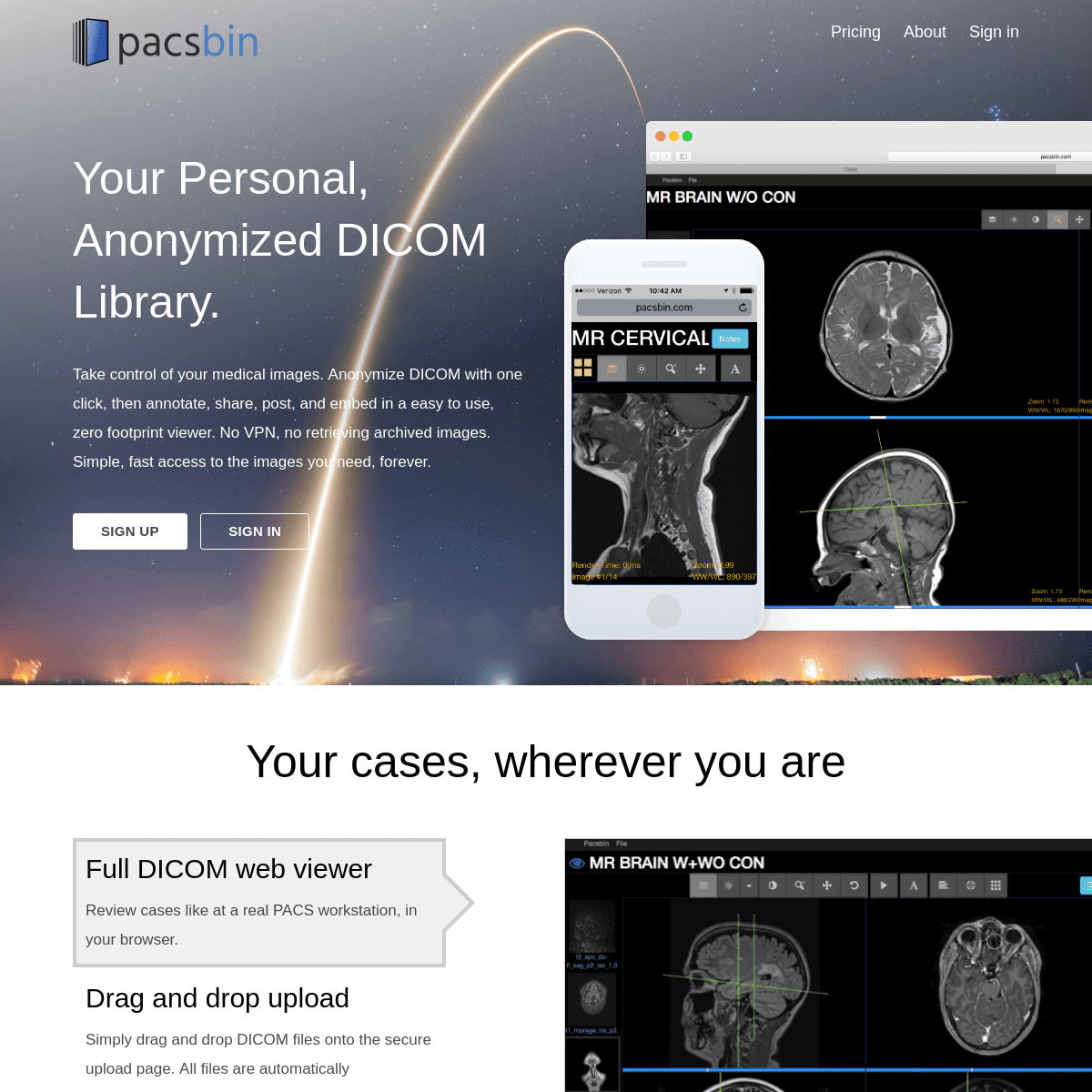
PACSBIN - YOUR PERSONAL CLOUD PACSSIGN INSIGN UPFLEISCHNER SOCIETYPRICINGABOUTSEND FEEDBACK Your Personal, Anonymized DICOM Library. Take control of your medical images. Anonymize DICOM with one click, then annotate, share, post, and embed in a easy to use, zero footprint viewer. No VPN, no retrieving archived images. Simple, fast access to the images you
need, forever.
SIGN IN - PACSBIN
Sign in with Facebook OR. EmailPACSBIN | ABOUT
Installing the Pacsbin proxy server where you work will allow you to securely look up patient data, even after exporting an anonymized study. This functionality requires that you are accessing Pacsbin from your work network, either while at work or using a remote access VPN. This ensures that no PHI is allowed outside the protected network. PACSBIN - YOUR PERSONAL CLOUD PACS Pro. $10 / month. $108 / year (save 10%!) Up to 500 studies. Share studies with colleagues. Create shared collections. Annotate studies with rich text and image links. Embed interactive DICOM studies in webpages. Create unlimited assessments.FLEISCHNER SOCIETY
Scroll tool: When active, click and drag on the image to scroll through the series of images. Adjust window/level: When active, click and drag on the image to adjust the brightness and contrast of the image, to bring out detail in different parts of the image. Common presets are available in the dropdown menu beside the button. Pan tool: When active, click and drag the image to move the image HOW TO READ ABDOMINAL CT: LABELED AND ORIGINAL IMAGES This is a CT of the Abdomen and Pelvis, Enterography protocol. This is a higher quality study than a standard CT. It is performed with a higher radiation dose and larger dose of IV contrast. The slice thickness is 2.5 mm. This provides a really excellent look at the large and small bowel enhancement and vasculature, and also the solidorgans.
33 Y/O MALE WITH APHASIA. STROKE EVAL. Presentation: 33 y/o male with aphasia. Stroke evaluation. Key findings: Patchy left multifocal MCA distribution foci of diffusion restriction (e.g., here and here) indicating acute ischemia/infarction.There is associated FLAIR siabnormality and correlative ADC mapping hypointensity (here and here).; Left frontal component contains small quantity petechial hemorrhage. 36 Y/O M WITH MARFAN SYNDROME AND CHEST PAIN. Presentation. 36 y/o M with Marfan syndrome and chest pain. Diagnosis. Type B aortic dissection. Key Findings. Intimal flap beginning just distal to the left subclavian artery and extending distal to the left renal artery, which arises from the false lumen.The celiac trunk, SMA, and right renal artery arise from the true lumen. The IMA is not wellvisualized.
BSGAR 1.4.05
BSGAR 1.4.05. Toggle Dropdown. Default. Soft Tissue (1) Bone (2) Brain(3) Stroke (4)
BSGAR 1.3.07
Toggle Dropdown. Slice location + Slice location - Acquisition time + Acquisition time - Image number + Image number - PACSBIN - YOUR PERSONAL CLOUD PACSSIGN INSIGN UPFLEISCHNER SOCIETYPRICINGABOUTSEND FEEDBACK Your Personal, Anonymized DICOM Library. Take control of your medical images. Anonymize DICOM with one click, then annotate, share, post, and embed in a easy to use, zero footprint viewer. No VPN, no retrieving archived images. Simple, fast access to the images youneed, forever.
SIGN IN - PACSBIN
Sign in with Facebook OR. EmailPACSBIN | ABOUT
Installing the Pacsbin proxy server where you work will allow you to securely look up patient data, even after exporting an anonymized study. This functionality requires that you are accessing Pacsbin from your work network, either while at work or using a remote access VPN. This ensures that no PHI is allowed outside the protected network. PACSBIN - YOUR PERSONAL CLOUD PACS Pro. $10 / month. $108 / year (save 10%!) Up to 500 studies. Share studies with colleagues. Create shared collections. Annotate studies with rich text and image links. Embed interactive DICOM studies in webpages. Create unlimited assessments.FLEISCHNER SOCIETY
Scroll tool: When active, click and drag on the image to scroll through the series of images. Adjust window/level: When active, click and drag on the image to adjust the brightness and contrast of the image, to bring out detail in different parts of the image. Common presets are available in the dropdown menu beside the button. Pan tool: When active, click and drag the image to move the image HOW TO READ ABDOMINAL CT: LABELED AND ORIGINAL IMAGES This is a CT of the Abdomen and Pelvis, Enterography protocol. This is a higher quality study than a standard CT. It is performed with a higher radiation dose and larger dose of IV contrast. The slice thickness is 2.5 mm. This provides a really excellent look at the large and small bowel enhancement and vasculature, and also the solidorgans.
33 Y/O MALE WITH APHASIA. STROKE EVAL. Presentation: 33 y/o male with aphasia. Stroke evaluation. Key findings: Patchy left multifocal MCA distribution foci of diffusion restriction (e.g., here and here) indicating acute ischemia/infarction.There is associated FLAIR siabnormality and correlative ADC mapping hypointensity (here and here).; Left frontal component contains small quantity petechial hemorrhage. 36 Y/O M WITH MARFAN SYNDROME AND CHEST PAIN. Presentation. 36 y/o M with Marfan syndrome and chest pain. Diagnosis. Type B aortic dissection. Key Findings. Intimal flap beginning just distal to the left subclavian artery and extending distal to the left renal artery, which arises from the false lumen.The celiac trunk, SMA, and right renal artery arise from the true lumen. The IMA is not wellvisualized.
BSGAR 1.4.05
BSGAR 1.4.05. Toggle Dropdown. Default. Soft Tissue (1) Bone (2) Brain(3) Stroke (4)
BSGAR 1.3.07
Toggle Dropdown. Slice location + Slice location - Acquisition time + Acquisition time - Image number + Image number -PACSBIN | ABOUT
Installing the Pacsbin proxy server where you work will allow you to securely look up patient data, even after exporting an anonymized study. This functionality requires that you are accessing Pacsbin from your work network, either while at work or using a remote access VPN. This ensures that no PHI is allowed outside the protected network.BSGAR 1.2.07
Image number -. Cor 3D MRCP NAV. COL:Cor 3D MRCP NAV. PANCREATIC DUCT slab MRCP. COR OBL 2D slab MRCP. Ax SSFSE (PURE) WATER: Ax LAVAFLEX. Render Time: Image #:BSGAR 1.2.13
Acquisition time -. Image number +. Image number -. undefined. Render Time: Image #: Zoom: WW/WC: Main duct IPMN with PDAC and chronic pancreatitis resected T2 N0 R0. GANGRENOUS CHOLECYSTITIS Hypoenhancement of the adjacent liver parenchyma due to inflammation. Common bile duct tapers rather abruptly on coronal reformats, which could suggest distal common bile duct stone. Diagnosis: Severe gangrenous cholecystitis. Management: Surgical.BSGAR 1.2.06
BSGAR 1.2.06. Toggle Dropdown. Default. Soft Tissue (1) Bone (2) Brain(3) Stroke (4)
TEACHING CT OF THE NECK Teaching CT of the neck Title hidden Toggle Dropdown. Default * Toggle Dropdown. Slice location + Slice location - Acquisition time +BSGAR 1.4.12
BSGAR 1.4.12. Toggle Dropdown. Default. Soft Tissue (1) Bone (2) Brain(3)
BSGAR 1.3.09
Toggle Dropdown. Slice location + Slice location - Acquisition time + Acquisition time - Image number + Image number -BSGAR 1.3.02
BSGAR 1.3.02. Toggle Dropdown. Default. Soft Tissue (1) Bone (2) Brain(3) Stroke (4)
BSGAR 1.3.01
BSGAR 1.3.01Title hidden. BSGAR 1.3.01. Toggle Dropdown. Default. Soft Tissue (1) Bone (2) Brain (3) Stroke (4) PACSBIN - YOUR PERSONAL CLOUD PACSSIGN INSIGN UPFLEISCHNER SOCIETYPRICINGABOUTSEND FEEDBACK Your Personal, Anonymized DICOM Library. Take control of your medical images. Anonymize DICOM with one click, then annotate, share, post, and embed in a easy to use, zero footprint viewer. No VPN, no retrieving archived images. Simple, fast access to the images youneed, forever.
SIGN IN - PACSBIN
Sign in with Facebook OR. EmailPACSBIN | ABOUT
Installing the Pacsbin proxy server where you work will allow you to securely look up patient data, even after exporting an anonymized study. This functionality requires that you are accessing Pacsbin from your work network, either while at work or using a remote access VPN. This ensures that no PHI is allowed outside the protected network. PACSBIN - YOUR PERSONAL CLOUD PACS Pro. $10 / month. $108 / year (save 10%!) Up to 500 studies. Share studies with colleagues. Create shared collections. Annotate studies with rich text and image links. Embed interactive DICOM studies in webpages. Create unlimited assessments. PACSBIN | INSTALL IMPAX BUTTON 1:05. Here are step by step instructions to install the button. Open any study in Impax. In the toolbar above the images, click the wrench button. (click to enlarge) Find and click the Wizards tab. (click to enlarge) You should see your button in the list of options. To add it to your toolbar, click and drag the button from the settings panelFLEISCHNER SOCIETY
Scroll tool: When active, click and drag on the image to scroll through the series of images. Adjust window/level: When active, click and drag on the image to adjust the brightness and contrast of the image, to bring out detail in different parts of the image. Common presets are available in the dropdown menu beside the button. Pan tool: When active, click and drag the image to move the image 33 Y/O MALE WITH APHASIA. STROKE EVAL. Presentation: 33 y/o male with aphasia. Stroke evaluation. Key findings: Patchy left multifocal MCA distribution foci of diffusion restriction (e.g., here and here) indicating acute ischemia/infarction.There is associated FLAIR siabnormality and correlative ADC mapping hypointensity (here and here).; Left frontal component contains small quantity petechial hemorrhage.BSGAR 1.2.10
Toggle Dropdown. Slice location + Slice location - Acquisition time + Acquisition time - Cystic fibrosis chronic pancreatitis missed PDACBSGAR 1.3.03
Toggle Dropdown. Slice location + Slice location - Acquisition time + Acquisition time - Image number + Image number -BSGAR 1.5.CHALLENGE
BSGAR 1.5.Challenge Title hidden Toggle Dropdown. Default * Toggle Dropdown. Slice location + Slice location - Acquisition time + PACSBIN - YOUR PERSONAL CLOUD PACSSIGN INSIGN UPFLEISCHNER SOCIETYPRICINGABOUTSEND FEEDBACK Your Personal, Anonymized DICOM Library. Take control of your medical images. Anonymize DICOM with one click, then annotate, share, post, and embed in a easy to use, zero footprint viewer. No VPN, no retrieving archived images. Simple, fast access to the images youneed, forever.
SIGN IN - PACSBIN
Sign in with Facebook OR. EmailPACSBIN | ABOUT
Installing the Pacsbin proxy server where you work will allow you to securely look up patient data, even after exporting an anonymized study. This functionality requires that you are accessing Pacsbin from your work network, either while at work or using a remote access VPN. This ensures that no PHI is allowed outside the protected network. PACSBIN - YOUR PERSONAL CLOUD PACS Pro. $10 / month. $108 / year (save 10%!) Up to 500 studies. Share studies with colleagues. Create shared collections. Annotate studies with rich text and image links. Embed interactive DICOM studies in webpages. Create unlimited assessments. PACSBIN | INSTALL IMPAX BUTTON 1:05. Here are step by step instructions to install the button. Open any study in Impax. In the toolbar above the images, click the wrench button. (click to enlarge) Find and click the Wizards tab. (click to enlarge) You should see your button in the list of options. To add it to your toolbar, click and drag the button from the settings panelFLEISCHNER SOCIETY
Scroll tool: When active, click and drag on the image to scroll through the series of images. Adjust window/level: When active, click and drag on the image to adjust the brightness and contrast of the image, to bring out detail in different parts of the image. Common presets are available in the dropdown menu beside the button. Pan tool: When active, click and drag the image to move the image 33 Y/O MALE WITH APHASIA. STROKE EVAL. Presentation: 33 y/o male with aphasia. Stroke evaluation. Key findings: Patchy left multifocal MCA distribution foci of diffusion restriction (e.g., here and here) indicating acute ischemia/infarction.There is associated FLAIR siabnormality and correlative ADC mapping hypointensity (here and here).; Left frontal component contains small quantity petechial hemorrhage.BSGAR 1.2.10
Toggle Dropdown. Slice location + Slice location - Acquisition time + Acquisition time - Cystic fibrosis chronic pancreatitis missed PDACBSGAR 1.3.03
Toggle Dropdown. Slice location + Slice location - Acquisition time + Acquisition time - Image number + Image number -BSGAR 1.5.CHALLENGE
BSGAR 1.5.Challenge Title hidden Toggle Dropdown. Default * Toggle Dropdown. Slice location + Slice location - Acquisition time +PACSBIN | ABOUT
Installing the Pacsbin proxy server where you work will allow you to securely look up patient data, even after exporting an anonymized study. This functionality requires that you are accessing Pacsbin from your work network, either while at work or using a remote access VPN. This ensures that no PHI is allowed outside the protected network.FLEISCHNER SOCIETY
Scroll tool: When active, click and drag on the image to scroll through the series of images. Adjust window/level: When active, click and drag on the image to adjust the brightness and contrast of the image, to bring out detail in different parts of the image. Common presets are available in the dropdown menu beside the button. Pan tool: When active, click and drag the image to move the image 33 Y/O MALE WITH APHASIA. STROKE EVAL. Presentation: 33 y/o male with aphasia. Stroke evaluation. Key findings: Patchy left multifocal MCA distribution foci of diffusion restriction (e.g., here and here) indicating acute ischemia/infarction.There is associated FLAIR siabnormality and correlative ADC mapping hypointensity (here and here).; Left frontal component contains small quantity petechial hemorrhage.BSGAR 1.2.07
Image number -. Cor 3D MRCP NAV. COL:Cor 3D MRCP NAV. PANCREATIC DUCT slab MRCP. COR OBL 2D slab MRCP. Ax SSFSE (PURE) WATER: Ax LAVAFLEX. Render Time: Image #:BSGAR 1.2.13
Acquisition time -. Image number +. Image number -. undefined. Render Time: Image #: Zoom: WW/WC: Main duct IPMN with PDAC and chronic pancreatitis resected T2 N0 R0.BSGAR 1.3.03
Toggle Dropdown. Slice location + Slice location - Acquisition time + Acquisition time - Image number + Image number -BSGAR 1.4.CHALLENGE
BSGAR 1.4.Challenge Title hidden Toggle Dropdown. Default * Toggle Dropdown. Slice location + Slice location - Acquisition time +BSGAR 1.4.08
Toggle Dropdown. Slice location + Slice location - Acquisition time + Acquisition time - Image number + Image number -BSGAR 1.3.02
BSGAR 1.3.02. Toggle Dropdown. Default. Soft Tissue (1) Bone (2) Brain(3) Stroke (4)
BSGAR 1.2.01
BSGAR 1.2.01Title hidden. BSGAR 1.2.01. Toggle Dropdown. Default. Soft Tissue (1) Bone (2) Brain (3) Stroke (4) PACSBIN - YOUR PERSONAL CLOUD PACSSIGN INSIGN UPFLEISCHNER SOCIETYPRICINGABOUTSEND FEEDBACK Your Personal, Anonymized DICOM Library. Take control of your medical images. Anonymize DICOM with one click, then annotate, share, post, and embed in a easy to use, zero footprint viewer. No VPN, no retrieving archived images. Simple, fast access to the images youneed, forever.
SIGN IN - PACSBIN
Sign in with Facebook OR. EmailPACSBIN | ABOUT
Installing the Pacsbin proxy server where you work will allow you to securely look up patient data, even after exporting an anonymized study. This functionality requires that you are accessing Pacsbin from your work network, either while at work or using a remote access VPN. This ensures that no PHI is allowed outside the protected network. PACSBIN - YOUR PERSONAL CLOUD PACS Pro. $10 / month. $108 / year (save 10%!) Up to 500 studies. Share studies with colleagues. Create shared collections. Annotate studies with rich text and image links. Embed interactive DICOM studies in webpages. Create unlimited assessments. PACSBIN | INSTALL IMPAX BUTTON 1:05. Here are step by step instructions to install the button. Open any study in Impax. In the toolbar above the images, click the wrench button. (click to enlarge) Find and click the Wizards tab. (click to enlarge) You should see your button in the list of options. To add it to your toolbar, click and drag the button from the settings panelFLEISCHNER SOCIETY
Scroll tool: When active, click and drag on the image to scroll through the series of images. Adjust window/level: When active, click and drag on the image to adjust the brightness and contrast of the image, to bring out detail in different parts of the image. Common presets are available in the dropdown menu beside the button. Pan tool: When active, click and drag the image to move the image 33 Y/O MALE WITH APHASIA. STROKE EVAL. Presentation: 33 y/o male with aphasia. Stroke evaluation. Key findings: Patchy left multifocal MCA distribution foci of diffusion restriction (e.g., here and here) indicating acute ischemia/infarction.There is associated FLAIR siabnormality and correlative ADC mapping hypointensity (here and here).; Left frontal component contains small quantity petechial hemorrhage.BSGAR 1.2.10
BSGAR 1.2.10. Toggle Dropdown. Default. Soft Tissue (1) Bone (2) Brain(3) Stroke (4)
BSGAR 1.3.03
Toggle Dropdown. Slice location + Slice location - Acquisition time + Acquisition time - Image number + Image number -BSGAR 1.2.01
BSGAR 1.2.01Title hidden. BSGAR 1.2.01. Toggle Dropdown. Default. Soft Tissue (1) Bone (2) Brain (3) Stroke (4)BSGAR 1.2.07
Image number -. Cor 3D MRCP NAV. COL:Cor 3D MRCP NAV. PANCREATIC DUCT slab MRCP. COR OBL 2D slab MRCP. Ax SSFSE (PURE) WATER: Ax LAVAFLEX. Render Time: Image #:BSGAR 1.2.12
BSGAR 1.2.12. Toggle Dropdown. Default. Soft Tissue (1) Bone (2) Brain(3)
BSGAR 1.4.03
BSGAR 1.4.03. Toggle Dropdown. Default. Soft Tissue (1) Bone (2) Brain(3)
BSGAR 1.3.04
Toggle Dropdown. Slice location + Slice location - Acquisition time + Acquisition time - Image number + Image number -BSGAR 1.4.CHALLENGE
BSGAR 1.4.Challenge Title hidden Toggle Dropdown. Default * Toggle Dropdown. Slice location + Slice location - Acquisition time + CASE 1: ACUTE APPENDICITIS CASE 1: Acute appendicitis. Presentation: Right lower quadrant pain, fever, leukocytosis. Click on the key findings to jump to the linkedimages!
BSGAR 1.2.13
Acquisition time -. Image number +. Image number -. undefined. Render Time: Image #: Zoom: WW/WC: Main duct IPMN with PDAC and chronic pancreatitis resected T2 N0 R0.BSGAR 1.3.02
BSGAR 1.3.02. Toggle Dropdown. Default. Soft Tissue (1) Bone (2) Brain(3) Stroke (4)
BSGAR 1.2.01
BSGAR 1.2.01Title hidden. BSGAR 1.2.01. Toggle Dropdown. Default. Soft Tissue (1) Bone (2) Brain (3) Stroke (4)BSGAR 1.2.02
Toggle Dropdown. Slice location + Slice location - Acquisition time + Acquisition time - Image number + Image number - PACSBIN - YOUR PERSONAL CLOUD PACSSIGN INSIGN UPFLEISCHNER SOCIETYPRICINGABOUTSEND FEEDBACK Your Personal, Anonymized DICOM Library. Take control of your medical images. Anonymize DICOM with one click, then annotate, share, post, and embed in a easy to use, zero footprint viewer.SIGN IN - PACSBIN
Sign in with Facebook OR. EmailPACSBIN | ABOUT
Patient Information Is patient information saved? Patient information, or protected health information (PHI) is never stored on Pacsbin.PACSBIN | ABOUT
About Pacsbin is a platform developed by radiologists as an attempt to make HIPAA compliant radiology teaching cases easier to create, view,and share.
SIGN IN - PACSBIN
Register new account. Sign in with Facebook OR PACSBIN - YOUR PERSONAL CLOUD PACS Up to 2000 studies; Share studies with colleagues Create shared collections Annotate studies with rich text and image links PACSBIN | INSTALL IMPAX BUTTONIMPAX PACSAGFA IMPAX PACSAGFA IMPAX 6 6IMPAX IMAGING SOFTWAREAGFA IMPAXAGFA IMPAX CLIENT Here are step by step instructions to install the button. Open any study in Impax. In the toolbar above the images, click the wrenchbutton.
FLEISCHNER SOCIETY
Scroll tool: When active, click and drag on the image to scroll through the series of images. Adjust window/level: When active, click and drag on the image to adjust the brightness and contrast of the image, to bring out detail in different parts of the image. Common presets are available in the dropdown menu beside the button. Pan tool: When active, click and drag the image to move the image HOW TO READ ABDOMINAL CT: LABELED AND ORIGINAL IMAGES Welcome to the teaching case for how to read a CT abdomen and pelvis! A few comments about the scan. This is a CT of the Abdomen and Pelvis, Enterography protocol TEACHING CT OF THE NECK Teaching CT of the neck Title hidden Toggle Dropdown. Default * Toggle Dropdown. Slice location + Slice location - Acquisition time + PACSBIN - YOUR PERSONAL CLOUD PACSSIGN INSIGN UPFLEISCHNER SOCIETYPRICINGABOUTSEND FEEDBACK Your Personal, Anonymized DICOM Library. Take control of your medical images. Anonymize DICOM with one click, then annotate, share, post, and embed in a easy to use, zero footprint viewer.SIGN IN - PACSBIN
Sign in with Facebook OR. EmailPACSBIN | ABOUT
Patient Information Is patient information saved? Patient information, or protected health information (PHI) is never stored on Pacsbin.PACSBIN | ABOUT
About Pacsbin is a platform developed by radiologists as an attempt to make HIPAA compliant radiology teaching cases easier to create, view,and share.
SIGN IN - PACSBIN
Register new account. Sign in with Facebook OR PACSBIN - YOUR PERSONAL CLOUD PACS Up to 2000 studies; Share studies with colleagues Create shared collections Annotate studies with rich text and image links PACSBIN | INSTALL IMPAX BUTTONIMPAX PACSAGFA IMPAX PACSAGFA IMPAX 6 6IMPAX IMAGING SOFTWAREAGFA IMPAXAGFA IMPAX CLIENT Here are step by step instructions to install the button. Open any study in Impax. In the toolbar above the images, click the wrenchbutton.
FLEISCHNER SOCIETY
Scroll tool: When active, click and drag on the image to scroll through the series of images. Adjust window/level: When active, click and drag on the image to adjust the brightness and contrast of the image, to bring out detail in different parts of the image. Common presets are available in the dropdown menu beside the button. Pan tool: When active, click and drag the image to move the image HOW TO READ ABDOMINAL CT: LABELED AND ORIGINAL IMAGES Welcome to the teaching case for how to read a CT abdomen and pelvis! A few comments about the scan. This is a CT of the Abdomen and Pelvis, Enterography protocol TEACHING CT OF THE NECK Teaching CT of the neck Title hidden Toggle Dropdown. Default * Toggle Dropdown. Slice location + Slice location - Acquisition time + GANGRENOUS CHOLECYSTITIS Presentation: 65 year-old female presenting with severe right upper quadrant pain. Key findings: Severe gallbladder dilatation with irregular enhancement. Irregular, discontinuous wall PACSBIN | SEND FEEDBACK Send Feedback Have a problem? A feature you'd like to suggest? Justwant to say Hi?
36 Y/O M WITH MARFAN SYNDROME AND CHEST PAIN. Presentation. 36 y/o M with Marfan syndrome and chest pain. Diagnosis. Type B aortic dissection. Key Findings. Intimal flap beginning just distal to the left subclavian artery and extending distal to the left renal artery, which arises from the false lumen.The celiac trunk, SMA, and right renal artery arise from the true lumen. The IMA is not wellvisualized.
SMALL BOWEL OBSTRUCTION Presentation: Nausea, vomiting, abdominal pain.History of small bowel resection. Key findings (GI tract in anatomic order): Stomach is moderately distended. Several loops of proximal small bowel are notdilated.
CASE 1: ACUTE APPENDICITIS Presentation: Right lower quadrant pain, fever, leukocytosis. Click on the key findings to jump to the linked images! Key findings: dilated, fluid filled appendix; appendicolith; inflammation/fluid 42-YEAR-OLD FEMALE WITH BACK PAIN AND WEAKNESS. HX OF IVDU. Presentation: 42-year-old female. Hx of IVDU, now with back pain and weakness. Key findings: Evidence of discitis-osteomyelitis involving the C4-C6 vertebral levels (T1 hypointense marrow signal spanning the discs).Here's a great example of how important the T1 is.; There's adjacent epidural abscess (best seen on diffusion-weighted imaging).This is really subtle/indistinguishable on the preBSGAR 1.2.10
Toggle Dropdown. Slice location + Slice location - Acquisition time + Acquisition time - Cystic fibrosis chronic pancreatitis missed PDACBSGAR 1.3.03
Toggle Dropdown. Slice location + Slice location - Acquisition time + Acquisition time - Image number + Image number -BSGAR 1.2.02
Toggle Dropdown. Slice location + Slice location - Acquisition time + Acquisition time - Image number + Image number -BSGAR 1.1.04
Toggle Dropdown. Slice location + Slice location - Acquisition time + Acquisition time - CBD stones on CT acute pancreatitis easy PACSBIN - YOUR PERSONAL CLOUD PACSSIGN INSIGN UPFLEISCHNER SOCIETYPRICINGABOUTSEND FEEDBACK Your Personal, Anonymized DICOM Library. Take control of your medical images. Anonymize DICOM with one click, then annotate, share, post, and embed in a easy to use, zero footprint viewer.SIGN IN - PACSBIN
Sign in with Facebook OR. EmailPACSBIN | ABOUT
Patient Information Is patient information saved? Patient information, or protected health information (PHI) is never stored on Pacsbin.PACSBIN | ABOUT
About Pacsbin is a platform developed by radiologists as an attempt to make HIPAA compliant radiology teaching cases easier to create, view,and share.
SIGN IN - PACSBIN
Register new account. Sign in with Facebook OR PACSBIN - YOUR PERSONAL CLOUD PACS Up to 2000 studies; Share studies with colleagues Create shared collections Annotate studies with rich text and image links PACSBIN | INSTALL IMPAX BUTTONIMPAX PACSAGFA IMPAX PACSAGFA IMPAX 6 6IMPAX IMAGING SOFTWAREAGFA IMPAXAGFA IMPAX CLIENT Here are step by step instructions to install the button. Open any study in Impax. In the toolbar above the images, click the wrenchbutton.
FLEISCHNER SOCIETY
Scroll tool: When active, click and drag on the image to scroll through the series of images. Adjust window/level: When active, click and drag on the image to adjust the brightness and contrast of the image, to bring out detail in different parts of the image. Common presets are available in the dropdown menu beside the button. Pan tool: When active, click and drag the image to move the image HOW TO READ ABDOMINAL CT: LABELED AND ORIGINAL IMAGES Welcome to the teaching case for how to read a CT abdomen and pelvis! A few comments about the scan. This is a CT of the Abdomen and Pelvis, Enterography protocol TEACHING CT OF THE NECK Teaching CT of the neck Title hidden Toggle Dropdown. Default * Toggle Dropdown. Slice location + Slice location - Acquisition time + PACSBIN - YOUR PERSONAL CLOUD PACSSIGN INSIGN UPFLEISCHNER SOCIETYPRICINGABOUTSEND FEEDBACK Your Personal, Anonymized DICOM Library. Take control of your medical images. Anonymize DICOM with one click, then annotate, share, post, and embed in a easy to use, zero footprint viewer.SIGN IN - PACSBIN
Sign in with Facebook OR. EmailPACSBIN | ABOUT
Patient Information Is patient information saved? Patient information, or protected health information (PHI) is never stored on Pacsbin.PACSBIN | ABOUT
About Pacsbin is a platform developed by radiologists as an attempt to make HIPAA compliant radiology teaching cases easier to create, view,and share.
SIGN IN - PACSBIN
Register new account. Sign in with Facebook OR PACSBIN - YOUR PERSONAL CLOUD PACS Up to 2000 studies; Share studies with colleagues Create shared collections Annotate studies with rich text and image links PACSBIN | INSTALL IMPAX BUTTONIMPAX PACSAGFA IMPAX PACSAGFA IMPAX 6 6IMPAX IMAGING SOFTWAREAGFA IMPAXAGFA IMPAX CLIENT Here are step by step instructions to install the button. Open any study in Impax. In the toolbar above the images, click the wrenchbutton.
FLEISCHNER SOCIETY
Scroll tool: When active, click and drag on the image to scroll through the series of images. Adjust window/level: When active, click and drag on the image to adjust the brightness and contrast of the image, to bring out detail in different parts of the image. Common presets are available in the dropdown menu beside the button. Pan tool: When active, click and drag the image to move the image HOW TO READ ABDOMINAL CT: LABELED AND ORIGINAL IMAGES Welcome to the teaching case for how to read a CT abdomen and pelvis! A few comments about the scan. This is a CT of the Abdomen and Pelvis, Enterography protocol TEACHING CT OF THE NECK Teaching CT of the neck Title hidden Toggle Dropdown. Default * Toggle Dropdown. Slice location + Slice location - Acquisition time + GANGRENOUS CHOLECYSTITIS Presentation: 65 year-old female presenting with severe right upper quadrant pain. Key findings: Severe gallbladder dilatation with irregular enhancement. Irregular, discontinuous wall PACSBIN | SEND FEEDBACK Send Feedback Have a problem? A feature you'd like to suggest? Justwant to say Hi?
36 Y/O M WITH MARFAN SYNDROME AND CHEST PAIN. Presentation. 36 y/o M with Marfan syndrome and chest pain. Diagnosis. Type B aortic dissection. Key Findings. Intimal flap beginning just distal to the left subclavian artery and extending distal to the left renal artery, which arises from the false lumen.The celiac trunk, SMA, and right renal artery arise from the true lumen. The IMA is not wellvisualized.
SMALL BOWEL OBSTRUCTION Presentation: Nausea, vomiting, abdominal pain.History of small bowel resection. Key findings (GI tract in anatomic order): Stomach is moderately distended. Several loops of proximal small bowel are notdilated.
CASE 1: ACUTE APPENDICITIS Presentation: Right lower quadrant pain, fever, leukocytosis. Click on the key findings to jump to the linked images! Key findings: dilated, fluid filled appendix; appendicolith; inflammation/fluid 42-YEAR-OLD FEMALE WITH BACK PAIN AND WEAKNESS. HX OF IVDU. Presentation: 42-year-old female. Hx of IVDU, now with back pain and weakness. Key findings: Evidence of discitis-osteomyelitis involving the C4-C6 vertebral levels (T1 hypointense marrow signal spanning the discs).Here's a great example of how important the T1 is.; There's adjacent epidural abscess (best seen on diffusion-weighted imaging).This is really subtle/indistinguishable on the preBSGAR 1.2.10
Toggle Dropdown. Slice location + Slice location - Acquisition time + Acquisition time - Cystic fibrosis chronic pancreatitis missed PDACBSGAR 1.3.03
Toggle Dropdown. Slice location + Slice location - Acquisition time + Acquisition time - Image number + Image number -BSGAR 1.2.02
Toggle Dropdown. Slice location + Slice location - Acquisition time + Acquisition time - Image number + Image number -BSGAR 1.1.04
Toggle Dropdown. Slice location + Slice location - Acquisition time + Acquisition time - CBD stones on CT acute pancreatitis easy Pricing About Sign in YOUR PERSONAL, ANONYMIZED DICOM LIBRARY. TAKE CONTROL OF YOUR MEDICAL IMAGES. ANONYMIZE DICOM WITH ONE CLICK, THEN ANNOTATE, SHARE, POST, AND EMBED IN A EASY TO USE, ZERO FOOTPRINT VIEWER. NO VPN, NO RETRIEVING ARCHIVED IMAGES. SIMPLE, FAST ACCESS TO THE IMAGES YOU NEED, FOREVER.Sign Up Sign In
YOUR CASES, WHEREVER YOU ARE FULL DICOM WEB VIEWER Review cases like at a real PACS workstation, in your browser. DRAG AND DROP UPLOAD Simply drag and drop DICOM files onto the secure upload page. All files are automatically anonymized and compressed without losingquality.
ONE CLICK PACS EXPORT Have a great case, but you're too busy to go through all the steps to save it? Click the Pacsbin button to start the secure export process and move on with your work.MOBILE READY
The Pacsbin zero-footprint web viewer gives you all the power of PACS anywhere you have a modern web browser.NO LOCK-IN
Switching jobs? Don't worry about losing your cases. Take them with you wherever you go, and download the anonymized full quality DICOMfiles at any time.
AUTO ANONYMIZATION
All studies added to Pacsbin are automatically anonymized, stripping sensitive information from DICOM headers and removing scanneddocuments.
START SHARING
Easily generate links to share studies with whomever you like, whether across the room or across the world. Share cases as unknowns, or highlight findings with custom annotations. REGISTER YOUR FREE ACCOUNTSign Up
�2016 Orion Medical Technologies, LLC Sign in Protected Health InformationPricing
Twitter Contact
Details
5