5
More Annotations


2


5
Favourite Annotations


4


5
Text
JANELIA RESEARCH CAMPUSOUR RESEARCHSUPPORT TEAMSOPEN SCIENCEYOU +
JANELIAABOUT USNEWS
Janelia Research Campus is HHMI’s pioneering research center in Ashburn, Virginia, where scientists pursue fundamental neuroscience and imaging in a uniquely innovative, collaborative atmosphere. FLYLIGHT | JANELIA RESEARCH CAMPUSTEAM MEMBERSTOOLS AND REAGENTSPROTOCOLSCOLLABORATIONS FlyLight plays a key role in this strategy by providing the bridge from electron-microscopic-level anatomical studies to behavioral and physiological functional studies. Cell-type-specific GAL4 drivers can be used to characterize the morphology of individual cells using the stochastic labeling methods we developed for this project. BIOLOGICAL TOOLS FOR 4D CELLULAR PHYSIOLOGY Organized by Luke Lavis (Janelia), Alison Tebo (Janelia) and Jordan Meier (NCI/NIH), this is the fourth in a series of planning workshops that will serve an important role in shaping a 15-lab, 15-year research program at Janelia Research Campus called “4D Cellular Physiology”.. The goal of 4DCP is to understand the function, structure and communication mechanisms of cells within and across JGCAMP8 CALCIUM INDICATORS The Looger Lab and the GENIE Project Team at HHMI Janelia have developed a new suite of jGCaMP8 calcium indicators, built on the GCaMP scaffold. The jGCaMP8 sensors have fast kinetics without compromising sensitivity, setting a new standard for in vivo imaging. MOUSELIGHT | JANELIA RESEARCH CAMPUS A major challenge in neuroscience is to relate the structure of neurons to their function. More than 100 years ago, Santiago Ramón y Cajal found that neurons are polarized cells with dendrites and axons, the input and output ends of neurons respectively. His discoveries were based on optical microscopy of histological preparations and single neuron reconstructions. LIPPINCOTT-SCHWARTZ LAB On a small scale, we are interested in the interplay between membrane-bound organelles, membrane-less organelles, cytoskeletal structures and metabolism as it relates to the organization and function of organ-specific cell types and their interactions. On a larger scale, we are trying to decipher how complex cellular behaviors characteristic of different cell types arise,FLYEM / HEMIBRAIN
The hemibrain connectome includes many of the brain areas that scientists are most interested in studying, such as circuits that control learning, memory, and key fly behaviors. The hemibrain connectome is the largest synaptic-level connectome ever reconstructed. It covers a large portion of the central fly brain, including the mushroom body and central complex circuitsSPLIT-GAL4 LINES
The Generation 1 GAL4 lines almost always express in more than one cell type. However, we found that it is possible to achieve cell-type specificity if expression is limited to the overlap in the expression patterns of two Generation 1 lines. The approach we prefer for carrying out such positive intersections is the split-GAL4 method (Luan et al. 2006; Pfeiffer et al, 2010).PAUL TILLBERG
tillbergp@janelia.hhmi.org. Fellow. node:field_people_current_research | entity_field. Paul completed a BS in the combined Electrical and Materials Engineering program at UC Berkeley in 2010. For his PhD, he focused on technology development for neuroscience at MIT. Using his background in polymeric materials, Paul conceived and co-developed aRAN DARSHAN
I received my bachelor's degree in physics and biology, as part of the Amirim and Etgar honors programs of the Hebrew University in Jerusalem. During these years I became interested in systems neuroscience and continued with pursuing a doctorate in computationaland
JANELIA RESEARCH CAMPUSOUR RESEARCHSUPPORT TEAMSOPEN SCIENCEYOU +JANELIAABOUT USNEWS
Janelia Research Campus is HHMI’s pioneering research center in Ashburn, Virginia, where scientists pursue fundamental neuroscience and imaging in a uniquely innovative, collaborative atmosphere. FLYLIGHT | JANELIA RESEARCH CAMPUSTEAM MEMBERSTOOLS AND REAGENTSPROTOCOLSCOLLABORATIONS FlyLight plays a key role in this strategy by providing the bridge from electron-microscopic-level anatomical studies to behavioral and physiological functional studies. Cell-type-specific GAL4 drivers can be used to characterize the morphology of individual cells using the stochastic labeling methods we developed for this project. BIOLOGICAL TOOLS FOR 4D CELLULAR PHYSIOLOGY Organized by Luke Lavis (Janelia), Alison Tebo (Janelia) and Jordan Meier (NCI/NIH), this is the fourth in a series of planning workshops that will serve an important role in shaping a 15-lab, 15-year research program at Janelia Research Campus called “4D Cellular Physiology”.. The goal of 4DCP is to understand the function, structure and communication mechanisms of cells within and across JGCAMP8 CALCIUM INDICATORS The Looger Lab and the GENIE Project Team at HHMI Janelia have developed a new suite of jGCaMP8 calcium indicators, built on the GCaMP scaffold. The jGCaMP8 sensors have fast kinetics without compromising sensitivity, setting a new standard for in vivo imaging. MOUSELIGHT | JANELIA RESEARCH CAMPUS A major challenge in neuroscience is to relate the structure of neurons to their function. More than 100 years ago, Santiago Ramón y Cajal found that neurons are polarized cells with dendrites and axons, the input and output ends of neurons respectively. His discoveries were based on optical microscopy of histological preparations and single neuron reconstructions. LIPPINCOTT-SCHWARTZ LAB On a small scale, we are interested in the interplay between membrane-bound organelles, membrane-less organelles, cytoskeletal structures and metabolism as it relates to the organization and function of organ-specific cell types and their interactions. On a larger scale, we are trying to decipher how complex cellular behaviors characteristic of different cell types arise,FLYEM / HEMIBRAIN
The hemibrain connectome includes many of the brain areas that scientists are most interested in studying, such as circuits that control learning, memory, and key fly behaviors. The hemibrain connectome is the largest synaptic-level connectome ever reconstructed. It covers a large portion of the central fly brain, including the mushroom body and central complex circuitsSPLIT-GAL4 LINES
The Generation 1 GAL4 lines almost always express in more than one cell type. However, we found that it is possible to achieve cell-type specificity if expression is limited to the overlap in the expression patterns of two Generation 1 lines. The approach we prefer for carrying out such positive intersections is the split-GAL4 method (Luan et al. 2006; Pfeiffer et al, 2010).PAUL TILLBERG
tillbergp@janelia.hhmi.org. Fellow. node:field_people_current_research | entity_field. Paul completed a BS in the combined Electrical and Materials Engineering program at UC Berkeley in 2010. For his PhD, he focused on technology development for neuroscience at MIT. Using his background in polymeric materials, Paul conceived and co-developed aRAN DARSHAN
I received my bachelor's degree in physics and biology, as part of the Amirim and Etgar honors programs of the Hebrew University in Jerusalem. During these years I became interested in systems neuroscience and continued with pursuing a doctorate in computationaland
VIRTUAL WORKSHOP PROGRAM The workshop "From Images to Knowledge with ImageJ & Friends" (I2K 2020) is fully virtual. Tutors and students live in various time zones around the globe. We therefore run almost all tutorials twice, once in the morning and once in the evening of the tutors' time-zone. Both sessions cover the same content, i.e. students participate in only onesession.
UNDERGRADUATE SCHOLARS PROGRAM The Janelia Undergraduate Scholars program is a 10-week summer program aimed at well-prepared, independent, and committed students with significant research experience. We accept undergraduates and masters students from around the world who have not committed to a PhD program. Participants must be actively enrolled as a student during the period of the Janelia Undergraduate JOINT GRADUATE PROGRAM The Janelia Joint Graduate Program is a fully-funded, collaborative PhD program for independent, committed graduate scholars in partnership with The Johns Hopkins University. Graduate scholars in the program spend their first year at the partner university and then reside at Janelia for the remaining years of the program to conduct their thesis research. MOUSELIGHT | JANELIA RESEARCH CAMPUS A major challenge in neuroscience is to relate the structure of neurons to their function. More than 100 years ago, Santiago Ramón y Cajal found that neurons are polarized cells with dendrites and axons, the input and output ends of neurons respectively. His discoveries were based on optical microscopy of histological preparations and single neuron reconstructions.FLYEM / HEMIBRAIN
The hemibrain connectome includes many of the brain areas that scientists are most interested in studying, such as circuits that control learning, memory, and key fly behaviors. The hemibrain connectome is the largest synaptic-level connectome ever reconstructed. It covers a large portion of the central fly brain, including the mushroom body and central complex circuits JGCAMP7 CALCIUM INDICATORS Plasmids available from Addgene Name Description Addgene reagent Reference pCMV-jGCaMP7s improved SNR, slow kinetics Plasmid Dana et al., 2019 pCMV-jGCaMP7f improved SNR, fast kinetics Plasmid Dana et al., 2019 pCMV-jGCaMP7b improved SNR, higher baseline fluorescence Plasmid Dana et al., 2019 pCMV-jGCaMP7c very low baseline fluorescence Plasmid Dana et al., 2019PAUL TILLBERG
tillbergp@janelia.hhmi.org. Fellow. node:field_people_current_research | entity_field. Paul completed a BS in the combined Electrical and Materials Engineering program at UC Berkeley in 2010. For his PhD, he focused on technology development for neuroscience at MIT. Using his background in polymeric materials, Paul conceived and co-developed aRAN DARSHAN
I received my bachelor's degree in physics and biology, as part of the Amirim and Etgar honors programs of the Hebrew University in Jerusalem. During these years I became interested in systems neuroscience and continued with pursuing a doctorate in computationaland
ALLA KARPOVA
I was very fortunate to grow up in the wonderful tradition of academic science in St. Petersburg, Russia, although it was not until college that I discovered biology. After high school, I had a unique opportunity to come to the United States to do my undergraduate studies at the University of Chicago. I loved math when I was a teenager and the University of Chicago’s amazingMARIUS PACHITARIU
I received my bachelor’s degree from Princeton University in mathematics, where I first discovered computational neuroscience through the guidance of Carlos Brody and Philip Holmes. I was fascinated by the complex analysis problems posed by neuroscience, but my training in mathematics was not enough to tackle them. I needed to learn machine learning, so I pursued a PhD at BIOLOGICAL TOOLS FOR 4D CELLULAR PHYSIOLOGY Organized by Luke Lavis (Janelia), Alison Tebo (Janelia) and Jordan Meier (NCI/NIH), this is the fourth in a series of planning workshops that will serve an important role in shaping a 15-lab, 15-year research program at Janelia Research Campus called “4D Cellular Physiology”.. The goal of 4DCP is to understand the function, structure and communication mechanisms of cells within and across FLYLIGHT | JANELIA RESEARCH CAMPUSTEAM MEMBERSTOOLS AND REAGENTSPROTOCOLSCOLLABORATIONS FlyLight plays a key role in this strategy by providing the bridge from electron-microscopic-level anatomical studies to behavioral and physiological functional studies. Cell-type-specific GAL4 drivers can be used to characterize the morphology of individual cells using the stochastic labeling methods we developed for this project. JGCAMP8 CALCIUM INDICATORS The Looger Lab and the GENIE Project Team at HHMI Janelia have developed a new suite of jGCaMP8 calcium indicators, built on the GCaMP scaffold. The jGCaMP8 sensors have fast kinetics without compromising sensitivity, setting a new standard for in vivo imaging.LEE (TZUMIN) LAB
Lee (Tzumin) Lab /. We are keen to determine the cellular complexity of the brain, to elucidate how numerous distinct neurons can derive from a limited number of progenitors, and to possibly reengineer the brain for understanding its structure, function and evolution. janelia7_blocks-janelia7_secondary_menu | block. More in this LabLanding Page.
SPLIT-GAL4 LINES
The Generation 1 GAL4 lines almost always express in more than one cell type. However, we found that it is possible to achieve cell-type specificity if expression is limited to the overlap in the expression patterns of two Generation 1 lines. The approach we prefer for carrying out such positive intersections is the split-GAL4 method (Luan et al. 2006; Pfeiffer et al, 2010).RAN DARSHAN
I received my bachelor's degree in physics and biology, as part of the Amirim and Etgar honors programs of the Hebrew University in Jerusalem. During these years I became interested in systems neuroscience and continued with pursuing a doctorate in computationaland
GWYNETH CARD
Gwyneth received her PhD. in Bioengineering from Caltech in Michael Dickinson's lab, where she studied Drosophila behavior and biomechanics. She has been a Group Leader at Janelia since 2010. Her lab is interested in simple perceptual decision-making and uses the Drosophila escape response as aSANDRO ROMANI
I joined Janelia in the fall of 2014 to study the dynamics of cortical circuits. My experience in the field of computational neuroscience started at the University of Rome La Sapienza. Under the supervision of Daniel Amit, covering the end of my Laurea studies and most of the PhD period, I witnessed unsupervised recurrent neural networks learn discrete sets of stimuli.PAUL TILLBERG
tillbergp@janelia.hhmi.org. Fellow. node:field_people_current_research | entity_field. Paul completed a BS in the combined Electrical and Materials Engineering program at UC Berkeley in 2010. For his PhD, he focused on technology development for neuroscience at MIT. Using his background in polymeric materials, Paul conceived and co-developed aKASPAR PODGORSKI
My goal is to peer into the brain and see how neurons process information. I studied Artificial Intelligence, Neuroscience, and Cognitive Science as an undergraduate at the University of Toronto. I was attracted by developments in optical physiology that allowed us to record atomic units of brain activity - synaptic currents and spikes - at ever-increasing scales. BIOLOGICAL TOOLS FOR 4D CELLULAR PHYSIOLOGY Organized by Luke Lavis (Janelia), Alison Tebo (Janelia) and Jordan Meier (NCI/NIH), this is the fourth in a series of planning workshops that will serve an important role in shaping a 15-lab, 15-year research program at Janelia Research Campus called “4D Cellular Physiology”.. The goal of 4DCP is to understand the function, structure and communication mechanisms of cells within and across FLYLIGHT | JANELIA RESEARCH CAMPUSTEAM MEMBERSTOOLS AND REAGENTSPROTOCOLSCOLLABORATIONS FlyLight plays a key role in this strategy by providing the bridge from electron-microscopic-level anatomical studies to behavioral and physiological functional studies. Cell-type-specific GAL4 drivers can be used to characterize the morphology of individual cells using the stochastic labeling methods we developed for this project. JGCAMP8 CALCIUM INDICATORS The Looger Lab and the GENIE Project Team at HHMI Janelia have developed a new suite of jGCaMP8 calcium indicators, built on the GCaMP scaffold. The jGCaMP8 sensors have fast kinetics without compromising sensitivity, setting a new standard for in vivo imaging.LEE (TZUMIN) LAB
Lee (Tzumin) Lab /. We are keen to determine the cellular complexity of the brain, to elucidate how numerous distinct neurons can derive from a limited number of progenitors, and to possibly reengineer the brain for understanding its structure, function and evolution. janelia7_blocks-janelia7_secondary_menu | block. More in this LabLanding Page.
SPLIT-GAL4 LINES
The Generation 1 GAL4 lines almost always express in more than one cell type. However, we found that it is possible to achieve cell-type specificity if expression is limited to the overlap in the expression patterns of two Generation 1 lines. The approach we prefer for carrying out such positive intersections is the split-GAL4 method (Luan et al. 2006; Pfeiffer et al, 2010).RAN DARSHAN
I received my bachelor's degree in physics and biology, as part of the Amirim and Etgar honors programs of the Hebrew University in Jerusalem. During these years I became interested in systems neuroscience and continued with pursuing a doctorate in computationaland
GWYNETH CARD
Gwyneth received her PhD. in Bioengineering from Caltech in Michael Dickinson's lab, where she studied Drosophila behavior and biomechanics. She has been a Group Leader at Janelia since 2010. Her lab is interested in simple perceptual decision-making and uses the Drosophila escape response as aSANDRO ROMANI
I joined Janelia in the fall of 2014 to study the dynamics of cortical circuits. My experience in the field of computational neuroscience started at the University of Rome La Sapienza. Under the supervision of Daniel Amit, covering the end of my Laurea studies and most of the PhD period, I witnessed unsupervised recurrent neural networks learn discrete sets of stimuli.PAUL TILLBERG
tillbergp@janelia.hhmi.org. Fellow. node:field_people_current_research | entity_field. Paul completed a BS in the combined Electrical and Materials Engineering program at UC Berkeley in 2010. For his PhD, he focused on technology development for neuroscience at MIT. Using his background in polymeric materials, Paul conceived and co-developed aKASPAR PODGORSKI
My goal is to peer into the brain and see how neurons process information. I studied Artificial Intelligence, Neuroscience, and Cognitive Science as an undergraduate at the University of Toronto. I was attracted by developments in optical physiology that allowed us to record atomic units of brain activity - synaptic currents and spikes - at ever-increasing scales. VIRTUAL WORKSHOP PROGRAM The workshop "From Images to Knowledge with ImageJ & Friends" (I2K 2020) is fully virtual. Tutors and students live in various time zones around the globe. We therefore run almost all tutorials twice, once in the morning and once in the evening of the tutors' time-zone. Both sessions cover the same content, i.e. students participate in only onesession.
LEE (TZUMIN) LAB
A “complete” cellular developmental map of adult Drosophila brain: The adult Drosophila cerebrum develops from about 100 pairs of NBs, each of which makes a characteristic lineage, a set of diverse neurons that form a NB clone. By stochastic clonal labeling, we have mapped the 100 or so NB clones in the adult Drosophila brain (Yu, Awasaki etal., 2013).
CELL BIOLOGICAL BASIS OF EPITHELIAL PHYSIOLOGY Organized by Jennifer Lippincott-Schwartz (Janelia), Michael Caplan (Yale) and Patrick Chitwood (Janelia), this is one in a series of planning workshops that will help identify where Janelia can make the most impact with a new 15-lab, 15-year research program.. In October, Janelia announced 4D Cellular Physiology as a new research area, complementing our first research area, Mechanistic JGCAMP7 CALCIUM INDICATORS Plasmids available from Addgene Name Description Addgene reagent Reference pCMV-jGCaMP7s improved SNR, slow kinetics Plasmid Dana et al., 2019 pCMV-jGCaMP7f improved SNR, fast kinetics Plasmid Dana et al., 2019 pCMV-jGCaMP7b improved SNR, higher baseline fluorescence Plasmid Dana et al., 2019 pCMV-jGCaMP7c very low baseline fluorescence Plasmid Dana et al., 2019 COLOR VISION: CIRCUITS AND BEHAVIOR Color Vision: Circuits and Behavior. Color vision plays a central role in many aspects of cognition and behavior, from social signaling and foraging, to spatial navigation and circadian rhythms. Recent advances in genetic and neuroanatomical methods are powering rapid progress in describing the circuits processing color information, which inSPLIT-GAL4 LINES
The Generation 1 GAL4 lines almost always express in more than one cell type. However, we found that it is possible to achieve cell-type specificity if expression is limited to the overlap in the expression patterns of two Generation 1 lines. The approach we prefer for carrying out such positive intersections is the split-GAL4 method (Luan et al. 2006; Pfeiffer et al, 2010).GWYNETH CARD
Gwyneth received her PhD. in Bioengineering from Caltech in Michael Dickinson's lab, where she studied Drosophila behavior and biomechanics. She has been a Group Leader at Janelia since 2010. Her lab is interested in simple perceptual decision-making and uses the Drosophila escape response as aPAUL TILLBERG
tillbergp@janelia.hhmi.org. Fellow. node:field_people_current_research | entity_field. Paul completed a BS in the combined Electrical and Materials Engineering program at UC Berkeley in 2010. For his PhD, he focused on technology development for neuroscience at MIT. Using his background in polymeric materials, Paul conceived and co-developed aJAMES ZHE LIU
Zhe Liu is a Group Leader at the Janelia Research Campus. He received a BS degree from Tsinghua University, China, and an MS degree in structural biology and biophysics Tsinghua University's School of Medicine. He received his PhD degree in molecular and cell biology from the University of California, Berkeley. At UC Berkeley he studied transcriptional regulation of stem cellMARIUS PACHITARIU
I received my bachelor’s degree from Princeton University in mathematics, where I first discovered computational neuroscience through the guidance of Carlos Brody and Philip Holmes. I was fascinated by the complex analysis problems posed by neuroscience, but my training in mathematics was not enough to tackle them. I needed to learn machine learning, so I pursued a PhD at BIOLOGICAL TOOLS FOR 4D CELLULAR PHYSIOLOGY Organized by Luke Lavis (Janelia), Alison Tebo (Janelia) and Jordan Meier (NCI/NIH), this is the fourth in a series of planning workshops that will serve an important role in shaping a 15-lab, 15-year research program at Janelia Research Campus called “4D Cellular Physiology”.. The goal of 4DCP is to understand the function, structure and communication mechanisms of cells within and across FLYLIGHT | JANELIA RESEARCH CAMPUSTEAM MEMBERSTOOLS AND REAGENTSPROTOCOLSCOLLABORATIONS FlyLight plays a key role in this strategy by providing the bridge from electron-microscopic-level anatomical studies to behavioral and physiological functional studies. Cell-type-specific GAL4 drivers can be used to characterize the morphology of individual cells using the stochastic labeling methods we developed for this project. JGCAMP8 CALCIUM INDICATORS The Looger Lab and the GENIE Project Team at HHMI Janelia have developed a new suite of jGCaMP8 calcium indicators, built on the GCaMP scaffold. The jGCaMP8 sensors have fast kinetics without compromising sensitivity, setting a new standard for in vivo imaging. JGCAMP7 CALCIUM INDICATORS Plasmids available from Addgene Name Description Addgene reagent Reference pCMV-jGCaMP7s improved SNR, slow kinetics Plasmid Dana et al., 2019 pCMV-jGCaMP7f improved SNR, fast kinetics Plasmid Dana et al., 2019 pCMV-jGCaMP7b improved SNR, higher baseline fluorescence Plasmid Dana et al., 2019 pCMV-jGCaMP7c very low baseline fluorescence Plasmid Dana et al., 2019SPLIT-GAL4 LINES
The Generation 1 GAL4 lines almost always express in more than one cell type. However, we found that it is possible to achieve cell-type specificity if expression is limited to the overlap in the expression patterns of two Generation 1 lines. The approach we prefer for carrying out such positive intersections is the split-GAL4 method (Luan et al. 2006; Pfeiffer et al, 2010).RAN DARSHAN
I received my bachelor's degree in physics and biology, as part of the Amirim and Etgar honors programs of the Hebrew University in Jerusalem. During these years I became interested in systems neuroscience and continued with pursuing a doctorate in computationaland
SANDRO ROMANI
I joined Janelia in the fall of 2014 to study the dynamics of cortical circuits. My experience in the field of computational neuroscience started at the University of Rome La Sapienza. Under the supervision of Daniel Amit, covering the end of my Laurea studies and most of the PhD period, I witnessed unsupervised recurrent neural networks learn discrete sets of stimuli.GWYNETH CARD
Gwyneth received her PhD. in Bioengineering from Caltech in Michael Dickinson's lab, where she studied Drosophila behavior and biomechanics. She has been a Group Leader at Janelia since 2010. Her lab is interested in simple perceptual decision-making and uses the Drosophila escape response as aPAUL TILLBERG
tillbergp@janelia.hhmi.org. Fellow. node:field_people_current_research | entity_field. Paul completed a BS in the combined Electrical and Materials Engineering program at UC Berkeley in 2010. For his PhD, he focused on technology development for neuroscience at MIT. Using his background in polymeric materials, Paul conceived and co-developed aKASPAR PODGORSKI
My goal is to peer into the brain and see how neurons process information. I studied Artificial Intelligence, Neuroscience, and Cognitive Science as an undergraduate at the University of Toronto. I was attracted by developments in optical physiology that allowed us to record atomic units of brain activity - synaptic currents and spikes - at ever-increasing scales. BIOLOGICAL TOOLS FOR 4D CELLULAR PHYSIOLOGY Organized by Luke Lavis (Janelia), Alison Tebo (Janelia) and Jordan Meier (NCI/NIH), this is the fourth in a series of planning workshops that will serve an important role in shaping a 15-lab, 15-year research program at Janelia Research Campus called “4D Cellular Physiology”.. The goal of 4DCP is to understand the function, structure and communication mechanisms of cells within and across FLYLIGHT | JANELIA RESEARCH CAMPUSTEAM MEMBERSTOOLS AND REAGENTSPROTOCOLSCOLLABORATIONS FlyLight plays a key role in this strategy by providing the bridge from electron-microscopic-level anatomical studies to behavioral and physiological functional studies. Cell-type-specific GAL4 drivers can be used to characterize the morphology of individual cells using the stochastic labeling methods we developed for this project. JGCAMP8 CALCIUM INDICATORS The Looger Lab and the GENIE Project Team at HHMI Janelia have developed a new suite of jGCaMP8 calcium indicators, built on the GCaMP scaffold. The jGCaMP8 sensors have fast kinetics without compromising sensitivity, setting a new standard for in vivo imaging. JGCAMP7 CALCIUM INDICATORS Plasmids available from Addgene Name Description Addgene reagent Reference pCMV-jGCaMP7s improved SNR, slow kinetics Plasmid Dana et al., 2019 pCMV-jGCaMP7f improved SNR, fast kinetics Plasmid Dana et al., 2019 pCMV-jGCaMP7b improved SNR, higher baseline fluorescence Plasmid Dana et al., 2019 pCMV-jGCaMP7c very low baseline fluorescence Plasmid Dana et al., 2019SPLIT-GAL4 LINES
The Generation 1 GAL4 lines almost always express in more than one cell type. However, we found that it is possible to achieve cell-type specificity if expression is limited to the overlap in the expression patterns of two Generation 1 lines. The approach we prefer for carrying out such positive intersections is the split-GAL4 method (Luan et al. 2006; Pfeiffer et al, 2010).RAN DARSHAN
I received my bachelor's degree in physics and biology, as part of the Amirim and Etgar honors programs of the Hebrew University in Jerusalem. During these years I became interested in systems neuroscience and continued with pursuing a doctorate in computationaland
SANDRO ROMANI
I joined Janelia in the fall of 2014 to study the dynamics of cortical circuits. My experience in the field of computational neuroscience started at the University of Rome La Sapienza. Under the supervision of Daniel Amit, covering the end of my Laurea studies and most of the PhD period, I witnessed unsupervised recurrent neural networks learn discrete sets of stimuli.GWYNETH CARD
Gwyneth received her PhD. in Bioengineering from Caltech in Michael Dickinson's lab, where she studied Drosophila behavior and biomechanics. She has been a Group Leader at Janelia since 2010. Her lab is interested in simple perceptual decision-making and uses the Drosophila escape response as aPAUL TILLBERG
tillbergp@janelia.hhmi.org. Fellow. node:field_people_current_research | entity_field. Paul completed a BS in the combined Electrical and Materials Engineering program at UC Berkeley in 2010. For his PhD, he focused on technology development for neuroscience at MIT. Using his background in polymeric materials, Paul conceived and co-developed aKASPAR PODGORSKI
My goal is to peer into the brain and see how neurons process information. I studied Artificial Intelligence, Neuroscience, and Cognitive Science as an undergraduate at the University of Toronto. I was attracted by developments in optical physiology that allowed us to record atomic units of brain activity - synaptic currents and spikes - at ever-increasing scales. CELL AND TISSUE CULTURE Our Cell and Tissue Culture Facility is a unique, consolidated facility providing expert maintenance of cell cultures with a high level of efficiency, competence, and safety for the research campus. This shared resource provides services for maintaining, plating, transfection and preparation of cells for imaging and otherapplications.
MOUSELIGHT | JANELIA RESEARCH CAMPUS A major challenge in neuroscience is to relate the structure of neurons to their function. More than 100 years ago, Santiago Ramón y Cajal found that neurons are polarized cells with dendrites and axons, the input and output ends of neurons respectively. His discoveries were based on optical microscopy of histological preparations and single neuron reconstructions. POSTDOCTORAL ASSOCIATES All labs at Janelia are internally funded at a level that allows scientists to pursue ambitious research programs. As a result, Janelia postdocs are free of grant writing and administrative responsibilities, which allows them to devote their energy and creativity to challenging, high-impact research. JGCAMP7 CALCIUM INDICATORS Plasmids available from Addgene Name Description Addgene reagent Reference pCMV-jGCaMP7s improved SNR, slow kinetics Plasmid Dana et al., 2019 pCMV-jGCaMP7f improved SNR, fast kinetics Plasmid Dana et al., 2019 pCMV-jGCaMP7b improved SNR, higher baseline fluorescence Plasmid Dana et al., 2019 pCMV-jGCaMP7c very low baseline fluorescence Plasmid Dana et al., 2019LEE (TZUMIN) LAB
A “complete” cellular developmental map of adult Drosophila brain: The adult Drosophila cerebrum develops from about 100 pairs of NBs, each of which makes a characteristic lineage, a set of diverse neurons that form a NB clone. By stochastic clonal labeling, we have mapped the 100 or so NB clones in the adult Drosophila brain (Yu, Awasaki etal., 2013).
DUDMAN LAB | JANELIA RESEARCH CAMPUS Published work Over the past few years we have focused on studying the circuits of the basal ganglia in the context of reward seeking behaviors in mice. Previous work has spanned a range of techniques and questions from behavioral measures of disease progression in human patients to the atomic structure of glutamate receptors. Data derived from: Google Scholar.GWYNETH CARD
Gwyneth received her PhD. in Bioengineering from Caltech in Michael Dickinson's lab, where she studied Drosophila behavior and biomechanics. She has been a Group Leader at Janelia since 2010. Her lab is interested in simple perceptual decision-making and uses the Drosophila escape response as aJAMES ZHE LIU
Zhe Liu is a Group Leader at the Janelia Research Campus. He received a BS degree from Tsinghua University, China, and an MS degree in structural biology and biophysics Tsinghua University's School of Medicine. He received his PhD degree in molecular and cell biology from the University of California, Berkeley. At UC Berkeley he studied transcriptional regulation of stem cellPAUL TILLBERG
tillbergp@janelia.hhmi.org. Fellow. node:field_people_current_research | entity_field. Paul completed a BS in the combined Electrical and Materials Engineering program at UC Berkeley in 2010. For his PhD, he focused on technology development for neuroscience at MIT. Using his background in polymeric materials, Paul conceived and co-developed aERIC SCHREITER
Eric received his undergraduate degree in chemistry from Truman State University in Missouri. During his Ph.D. work in Biological Chemistry with Catherine L. Drennan at the Massachusetts Institute of Technology, he worked on the molecular mechanism of nickel ion homeostasis in bacteria by solving x-ray crystal structures of the nickel-responsive transcription factor NikR. BIOLOGICAL TOOLS FOR 4D CELLULAR PHYSIOLOGY Organized by Luke Lavis (Janelia), Alison Tebo (Janelia) and Jordan Meier (NCI/NIH), this is the fourth in a series of planning workshops that will serve an important role in shaping a 15-lab, 15-year research program at Janelia Research Campus called “4D Cellular Physiology”.. The goal of 4DCP is to understand the function, structure and communication mechanisms of cells within and across FLYLIGHT | JANELIA RESEARCH CAMPUSTEAM MEMBERSTOOLS AND REAGENTSPROTOCOLSCOLLABORATIONS FlyLight plays a key role in this strategy by providing the bridge from electron-microscopic-level anatomical studies to behavioral and physiological functional studies. Cell-type-specific GAL4 drivers can be used to characterize the morphology of individual cells using the stochastic labeling methods we developed for this project. JGCAMP8 CALCIUM INDICATORS The Looger Lab and the GENIE Project Team at HHMI Janelia have developed a new suite of jGCaMP8 calcium indicators, built on the GCaMP scaffold. The jGCaMP8 sensors have fast kinetics without compromising sensitivity, setting a new standard for in vivo imaging. JGCAMP7 CALCIUM INDICATORS Plasmids available from Addgene Name Description Addgene reagent Reference pCMV-jGCaMP7s improved SNR, slow kinetics Plasmid Dana et al., 2019 pCMV-jGCaMP7f improved SNR, fast kinetics Plasmid Dana et al., 2019 pCMV-jGCaMP7b improved SNR, higher baseline fluorescence Plasmid Dana et al., 2019 pCMV-jGCaMP7c very low baseline fluorescence Plasmid Dana et al., 2019SPLIT-GAL4 LINES
The Generation 1 GAL4 lines almost always express in more than one cell type. However, we found that it is possible to achieve cell-type specificity if expression is limited to the overlap in the expression patterns of two Generation 1 lines. The approach we prefer for carrying out such positive intersections is the split-GAL4 method (Luan et al. 2006; Pfeiffer et al, 2010).RAN DARSHAN
I received my bachelor's degree in physics and biology, as part of the Amirim and Etgar honors programs of the Hebrew University in Jerusalem. During these years I became interested in systems neuroscience and continued with pursuing a doctorate in computationaland
SANDRO ROMANI
I joined Janelia in the fall of 2014 to study the dynamics of cortical circuits. My experience in the field of computational neuroscience started at the University of Rome La Sapienza. Under the supervision of Daniel Amit, covering the end of my Laurea studies and most of the PhD period, I witnessed unsupervised recurrent neural networks learn discrete sets of stimuli.GWYNETH CARD
Gwyneth received her PhD. in Bioengineering from Caltech in Michael Dickinson's lab, where she studied Drosophila behavior and biomechanics. She has been a Group Leader at Janelia since 2010. Her lab is interested in simple perceptual decision-making and uses the Drosophila escape response as aPAUL TILLBERG
tillbergp@janelia.hhmi.org. Fellow. node:field_people_current_research | entity_field. Paul completed a BS in the combined Electrical and Materials Engineering program at UC Berkeley in 2010. For his PhD, he focused on technology development for neuroscience at MIT. Using his background in polymeric materials, Paul conceived and co-developed aKASPAR PODGORSKI
My goal is to peer into the brain and see how neurons process information. I studied Artificial Intelligence, Neuroscience, and Cognitive Science as an undergraduate at the University of Toronto. I was attracted by developments in optical physiology that allowed us to record atomic units of brain activity - synaptic currents and spikes - at ever-increasing scales. BIOLOGICAL TOOLS FOR 4D CELLULAR PHYSIOLOGY Organized by Luke Lavis (Janelia), Alison Tebo (Janelia) and Jordan Meier (NCI/NIH), this is the fourth in a series of planning workshops that will serve an important role in shaping a 15-lab, 15-year research program at Janelia Research Campus called “4D Cellular Physiology”.. The goal of 4DCP is to understand the function, structure and communication mechanisms of cells within and across FLYLIGHT | JANELIA RESEARCH CAMPUSTEAM MEMBERSTOOLS AND REAGENTSPROTOCOLSCOLLABORATIONS FlyLight plays a key role in this strategy by providing the bridge from electron-microscopic-level anatomical studies to behavioral and physiological functional studies. Cell-type-specific GAL4 drivers can be used to characterize the morphology of individual cells using the stochastic labeling methods we developed for this project. JGCAMP8 CALCIUM INDICATORS The Looger Lab and the GENIE Project Team at HHMI Janelia have developed a new suite of jGCaMP8 calcium indicators, built on the GCaMP scaffold. The jGCaMP8 sensors have fast kinetics without compromising sensitivity, setting a new standard for in vivo imaging. JGCAMP7 CALCIUM INDICATORS Plasmids available from Addgene Name Description Addgene reagent Reference pCMV-jGCaMP7s improved SNR, slow kinetics Plasmid Dana et al., 2019 pCMV-jGCaMP7f improved SNR, fast kinetics Plasmid Dana et al., 2019 pCMV-jGCaMP7b improved SNR, higher baseline fluorescence Plasmid Dana et al., 2019 pCMV-jGCaMP7c very low baseline fluorescence Plasmid Dana et al., 2019SPLIT-GAL4 LINES
The Generation 1 GAL4 lines almost always express in more than one cell type. However, we found that it is possible to achieve cell-type specificity if expression is limited to the overlap in the expression patterns of two Generation 1 lines. The approach we prefer for carrying out such positive intersections is the split-GAL4 method (Luan et al. 2006; Pfeiffer et al, 2010).RAN DARSHAN
I received my bachelor's degree in physics and biology, as part of the Amirim and Etgar honors programs of the Hebrew University in Jerusalem. During these years I became interested in systems neuroscience and continued with pursuing a doctorate in computationaland
SANDRO ROMANI
I joined Janelia in the fall of 2014 to study the dynamics of cortical circuits. My experience in the field of computational neuroscience started at the University of Rome La Sapienza. Under the supervision of Daniel Amit, covering the end of my Laurea studies and most of the PhD period, I witnessed unsupervised recurrent neural networks learn discrete sets of stimuli.GWYNETH CARD
Gwyneth received her PhD. in Bioengineering from Caltech in Michael Dickinson's lab, where she studied Drosophila behavior and biomechanics. She has been a Group Leader at Janelia since 2010. Her lab is interested in simple perceptual decision-making and uses the Drosophila escape response as aPAUL TILLBERG
tillbergp@janelia.hhmi.org. Fellow. node:field_people_current_research | entity_field. Paul completed a BS in the combined Electrical and Materials Engineering program at UC Berkeley in 2010. For his PhD, he focused on technology development for neuroscience at MIT. Using his background in polymeric materials, Paul conceived and co-developed aKASPAR PODGORSKI
My goal is to peer into the brain and see how neurons process information. I studied Artificial Intelligence, Neuroscience, and Cognitive Science as an undergraduate at the University of Toronto. I was attracted by developments in optical physiology that allowed us to record atomic units of brain activity - synaptic currents and spikes - at ever-increasing scales. CELL AND TISSUE CULTURE Our Cell and Tissue Culture Facility is a unique, consolidated facility providing expert maintenance of cell cultures with a high level of efficiency, competence, and safety for the research campus. This shared resource provides services for maintaining, plating, transfection and preparation of cells for imaging and otherapplications.
MOUSELIGHT | JANELIA RESEARCH CAMPUS A major challenge in neuroscience is to relate the structure of neurons to their function. More than 100 years ago, Santiago Ramón y Cajal found that neurons are polarized cells with dendrites and axons, the input and output ends of neurons respectively. His discoveries were based on optical microscopy of histological preparations and single neuron reconstructions. POSTDOCTORAL ASSOCIATES All labs at Janelia are internally funded at a level that allows scientists to pursue ambitious research programs. As a result, Janelia postdocs are free of grant writing and administrative responsibilities, which allows them to devote their energy and creativity to challenging, high-impact research. JGCAMP7 CALCIUM INDICATORS Plasmids available from Addgene Name Description Addgene reagent Reference pCMV-jGCaMP7s improved SNR, slow kinetics Plasmid Dana et al., 2019 pCMV-jGCaMP7f improved SNR, fast kinetics Plasmid Dana et al., 2019 pCMV-jGCaMP7b improved SNR, higher baseline fluorescence Plasmid Dana et al., 2019 pCMV-jGCaMP7c very low baseline fluorescence Plasmid Dana et al., 2019LEE (TZUMIN) LAB
A “complete” cellular developmental map of adult Drosophila brain: The adult Drosophila cerebrum develops from about 100 pairs of NBs, each of which makes a characteristic lineage, a set of diverse neurons that form a NB clone. By stochastic clonal labeling, we have mapped the 100 or so NB clones in the adult Drosophila brain (Yu, Awasaki etal., 2013).
DUDMAN LAB | JANELIA RESEARCH CAMPUS Published work Over the past few years we have focused on studying the circuits of the basal ganglia in the context of reward seeking behaviors in mice. Previous work has spanned a range of techniques and questions from behavioral measures of disease progression in human patients to the atomic structure of glutamate receptors. Data derived from: Google Scholar.GWYNETH CARD
Gwyneth received her PhD. in Bioengineering from Caltech in Michael Dickinson's lab, where she studied Drosophila behavior and biomechanics. She has been a Group Leader at Janelia since 2010. Her lab is interested in simple perceptual decision-making and uses the Drosophila escape response as aJAMES ZHE LIU
Zhe Liu is a Group Leader at the Janelia Research Campus. He received a BS degree from Tsinghua University, China, and an MS degree in structural biology and biophysics Tsinghua University's School of Medicine. He received his PhD degree in molecular and cell biology from the University of California, Berkeley. At UC Berkeley he studied transcriptional regulation of stem cellPAUL TILLBERG
tillbergp@janelia.hhmi.org. Fellow. node:field_people_current_research | entity_field. Paul completed a BS in the combined Electrical and Materials Engineering program at UC Berkeley in 2010. For his PhD, he focused on technology development for neuroscience at MIT. Using his background in polymeric materials, Paul conceived and co-developed aERIC SCHREITER
Eric received his undergraduate degree in chemistry from Truman State University in Missouri. During his Ph.D. work in Biological Chemistry with Catherine L. Drennan at the Massachusetts Institute of Technology, he worked on the molecular mechanism of nickel ion homeostasis in bacteria by solving x-ray crystal structures of the nickel-responsive transcription factor NikR. Skip to main content MAIN MENU (MOBILE)- BLOCKMENU
* Our Research
* Overview
* Our Labs
* Project Teams
* Former Labs
* Research Area Competition* Support Teams
* Overview
* Anatomy and Histology * Cell and Tissue Culture * Connectome Annotation * Cryo-Electron Microscopy * Drosophila Resources * Electron Microscopy * Gene Targeting and Transgenics * Janelia Experimental Technology* Light Microscopy
* Media Prep
* Molecular Biology
* Project Technical Resources * Quantitative Genomics * Scientific Computing Software * Scientific Computing Systems* Viral Tools
* Vivarium
* Open Science
* Overview
* Tools & Innovations * Advanced Imaging Center* You + Janelia
* Overview
* Careers
* Students and Postdocs * Conferences and Workshops * Visiting Scientists* Dialogues
* About Us
* Overview
* History
* Leadership
* Intellectual Life at Janelia * Research Facilities* Campus Amenities
* Janelia in the Community* News
* Careers
* Conferences
* People
* Publications
* Research Groups
Labs:
View All Labs at JaneliaLabs A-C
Ahrens Lab
Aso Lab
Betzig Lab
Beyene Lab
Branson Lab
Card Lab
Clapham Lab
Labs D-H
Darshan Lab
Dickson Lab
Dudman Lab
Fitzgerald Lab
Funke Lab
Grigorieff Lab
Hantman Lab
Harris Lab
Heberlein Lab
Hermundstad Lab
Hess Lab
Huston Lab
Labs I-L
Jayaraman Lab
Karpova Lab
Keleman Lab
Keller Lab
Koyama Lab
Lavis Lab
Lee (Albert) Lab
Lee (Tzumin) Lab
Leonardo Lab
Lippincott-Schwartz LabLiu Lab
Looger Lab
Labs M-S
O'Shea Lab
Pachitariu Lab
Podgorski Lab
Reiser Lab
Romani Lab
Rubin Lab
Ryan Lab
Saalfeld Lab
Scheffer Lab
Schreiter Lab
Singer Lab
Spruston Lab
Stern Lab
Sternson Lab
Svoboda Lab
Labs T-Z
Tillberg Lab
Truman Lab
Turaga Lab
Turner Lab
Ahrens Lab
Aso Lab
Betzig Lab
Beyene Lab
Branson Lab
Card Lab
Clapham Lab
Darshan Lab
Dickson Lab
Dudman Lab
Fitzgerald Lab
Funke Lab
Grigorieff Lab
Hantman Lab
Harris Lab
Heberlein Lab
Hermundstad Lab
Hess Lab
Huston Lab
Jayaraman Lab
Karpova Lab
Keleman Lab
Keller Lab
Koyama Lab
Lavis Lab
Lee (Albert) Lab
Lee (Tzumin) Lab
Leonardo Lab
Lippincott-Schwartz LabLiu Lab
Looger Lab
O'Shea Lab
Pachitariu Lab
Podgorski Lab
Reiser Lab
Romani Lab
Rubin Lab
Ryan Lab
Saalfeld Lab
Scheffer Lab
Schreiter Lab
Singer Lab
Spruston Lab
Stern Lab
Sternson Lab
Svoboda Lab
Tillberg Lab
Truman Lab
Turaga Lab
Turner Lab
Project Teams:
COSEM
Fly Descending InterneuronFlyEM
FlyLight
GENIE
MouseLight
MAIN MENU - BLOCK
* News
* Careers
* Conferences
* People
* Publications
* Research Groups
* Our Research
* Support Teams
* Open Science
* You + Janelia
* About Us
Labs:
View All Labs at JaneliaAhrens Lab
Aso Lab
Betzig Lab
Beyene Lab
Branson Lab
Card Lab
Clapham Lab
Darshan Lab
Dickson Lab
Dudman Lab
Fitzgerald Lab
Funke Lab
Grigorieff Lab
Hantman Lab
Harris Lab
Heberlein Lab
Hermundstad Lab
Hess Lab
Huston Lab
Jayaraman Lab
Karpova Lab
Keleman Lab
Keller Lab
Koyama Lab
Lavis Lab
Lee (Albert) Lab
Lee (Tzumin) Lab
Leonardo Lab
Lippincott-Schwartz LabLiu Lab
Looger Lab
O'Shea Lab
Pachitariu Lab
Podgorski Lab
Reiser Lab
Romani Lab
Rubin Lab
Ryan Lab
Saalfeld Lab
Scheffer Lab
Schreiter Lab
Singer Lab
Spruston Lab
Stern Lab
Sternson Lab
Svoboda Lab
Tillberg Lab
Truman Lab
Turaga Lab
Turner Lab
Project Teams:
COSEM
Fly Descending InterneuronFlyEM
FlyLight
GENIE
MouseLight
* Overview
* Our Labs
* Project Teams
* Former Labs
* Research Area Competition* Overview
* Anatomy and Histology * Cell and Tissue Culture * Connectome Annotation * Cryo-Electron Microscopy * Drosophila Resources * Electron Microscopy * Gene Targeting and Transgenics * Janelia Experimental Technology* Light Microscopy
* Media Prep
* Molecular Biology
* Project Technical Resources * Quantitative Genomics * Scientific Computing Software * Scientific Computing Systems* Viral Tools
* Vivarium
* Overview
* Tools & Innovations * Advanced Imaging Center* Overview
* Careers
* Students and Postdocs * Conferences and Workshops * Visiting Scientists* Dialogues
* Overview
* History
* Leadership
* Intellectual Life at Janelia * Research Facilities* Campus Amenities
* Janelia in the CommunityHOMEPAGE
homepage_slideshow | views Small labs, big science. janelia_helper-janelia7_publication_list | blockPUBLICATIONS
Lavis Lab Schreiter Lab Molecular Biology Cell and Tissue CultureViral Tools
Bright and tunable far-red chemigenetic indicators. Lippincott-Schwartz Lab Betzig Lab Hess Lab Janelia ExperimentalTechnology
Correlative three-dimensional super-resolution and block-face electron microscopy of whole vitreously frozen cells. Branson Lab Hantman Lab Anatomy and Histology Scientific Computing Systems Janelia Experimental Technology Cortical pattern generation during dexterous movement isinput-driven.
Looger Lab Drosophila Resources Quantitative Genomics The neuropeptide Drosulfakinin regulates social isolation-induced aggression in Drosophila.Harris Lab
Neuropixels data-acquisition system: A scalable platform for parallel recording of 10 000+ electrophysiological signals. Turner Lab Podgorski Lab Two-photon imaging with silicon photomultipliers. Romani Lab Reiser Lab The computation of directional selectivity in the OFF motion pathway. Liu Lab Lippincott-Schwartz Lab Phase separation of YAP reorganizes genome topology for long-term YAP target gene expression. janelia_helper-janelia7_view_all_publications | block View All Publications janelia7_blocks-janelia7_featured_stories_grid | blockFEATURED
Announcement
04/09/2020 | Janelia’s commitment to research continues even as our campus operations are reduced due to COVID-19. Here are some ways that Janelians are sharing their time, expertise, and resources in thismoment of need.
News
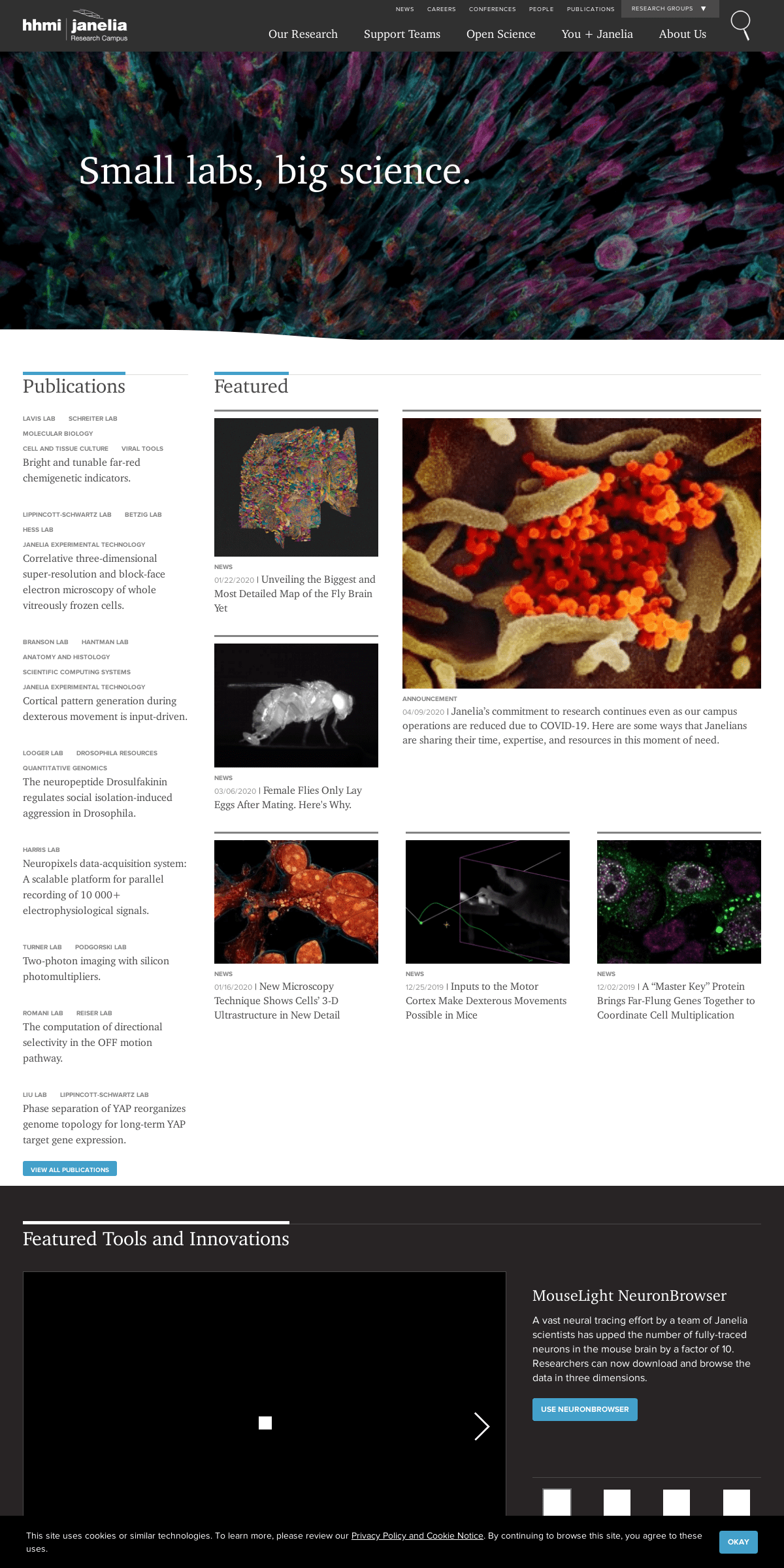
01/22/2020 | Unveiling the Biggest and Most Detailed Map of the FlyBrain Yet
News
03/06/2020 | Female Flies Only Lay Eggs After Mating. Here's Why.Announcement
04/09/2020 | Janelia’s commitment to research continues even as our campus operations are reduced due to COVID-19. Here are some ways that Janelians are sharing their time, expertise, and resources in thismoment of need.
News
01/16/2020 | New Microscopy Technique Shows Cells’ 3-D Ultrastructure in New DetailNews
12/25/2019 | Inputs to the Motor Cortex Make Dexterous MovementsPossible in Mice
News
12/02/2019 | A “Master Key” Protein Brings Far-Flung Genes Together to Coordinate Cell Multiplication janelia7_blocks-janelia7_featured_stories2_grid | blockcustom | custom
janelia7_blocks-janelia7_homepage_promo | block node:field_gallery_title | entity_field Featured Tools and Innovations janelia7_blocks-janelia7_media_gallery | blockLEFT RIGHT
MouseLight NeuronBrowser A vast neural tracing effort by a team of Janelia scientists has upped the number of fully-traced neurons in the mouse brain by a factor of 10. Researchers can now download and browse the data in threedimensions.
Use NeuronBrowser
Hemidriver Drosophila Lines ("Split-halves") These split-GAL4 driver Drosophila lines allow the generation of cell-type specific gene expression. They are freely available through the Bloomington Stockcenter through a large coordinated effort from Janelia Drosophila Resources and the National Institutes of Health.Learn More
Janelia Fluor Dyes
These bright and photostable fluorescent labels enable imaging and tracking of individual molecules inside living cells.Learn More
GCaMP6
The GCaMP6 family of GECIs is a collection of green fluorescent indicator proteins that facilitate the measurement of synaptic calcium signals, enabling reliable detection of neuronal responses in vivo. The next-generation GCaMP6s, GCaMP6m, and GCaMP6f sensors offer increased sensitivity (single action potential detection in vivo) andimproved kinetics.
Learn More
CaMPARI
This photoconvertible protein enables imaging of the calcium activity history of large areas and populations of cells. It is based on EosFP, a fluorescent protein that shows emission changes from green to red upon irradiation with UV light. It is not restricted to the field of view of a microscope, as during real-timecalcium imaging.
Learn More
Lattice Light Sheet Microscope This microscope uses a Bessel beam to illuminate a sample with light sheets that are sufficiently thin to achieve isotropic resolution high-speed 3D fluorescent imaging. The technology can visualize cellular processes in spatiotemporal detail not attainable with other fluorescent microscopy techniques.Learn More
LEFT RIGHT
* About Janelia
* Careers
* Get Directions
* Privacy Policy & Cookie Notice* News
* HHMI.org
571.209.4000
Find us:
19700 Helix Drive, Ashburn, Virginia 20147 19700 Helix Drive, Ashburn, Virginia 20147571.209.4000
Contact Us
Copyright © 2020 Howard Hughes Medical InstituteLog In
Contact Us
This site uses cookies or similar technologies. To learn more, please review our Privacy Policy and Cookie Notice . By continuing to browse this site, you agree to these uses.Okay
Details
2