5
More Annotations


6


3
Favourite Annotations


1


2
Text
HOME | ACLAND'S VIDEO ATLAS OF HUMAN ANATOMYEXAMSA-Z INDEXABOUTGLOSSARYVOLUME 1: THE UPPER EXTREMITY Real Movement. Exquisite Dissections. Acland's Video Atlas of Human Anatomy contains nearly 330 videos of real human anatomic specimens in their natural colors, including 5 new, groundbreaking videos of the inner ear. Dr. Robert Acland presents moving structures—muscles, tendons, and joints—making the same movements that they make in
life.
EXAMS | ACLAND'S VIDEO ATLAS OF HUMAN ANATOMY In order to use all the great features Acland's Video Atlas of Human Anatomy has to offer, you must create a personal account. Sign Up Now! ACLAND'S VIDEO ATLAS OF HUMAN ANATOMY We are unable to redeem your access code. Please try again anothertime.
VOCAL LIGAMENTS, VOCAL OPENING (2.29) Now that we’ve looked at the skeleton of the larynx, it’s time to get acquainted with the vocal ligament and the vocal opening. To see the vocal ligament, we’ll look at a specimen in which the lamina of the thyroid cartilage has been removed on the right side. BRAIN: INITIAL OVERVIEW (0.46) In this section we'll first take a brief look at the shape of the brain and its main parts, then we'll look at the cavity that contains it and the layers of tissue that surround it, then we'll return to the brain itself and look at it in more detail. “GETTING STARTED” USER GUIDE An expanded table of contents for each video is listed in the panel on the left side of the video window, allowing you to Jump To specific parts of the video. You can LONG AND SHORT TOE EXTENSOR MUSCLES (3.24) Now we’ll look at the muscles which produce movement of the toes. We’ll look at the extensor muscles first. There are two long extensors to the toes, and two short ones. URETER | ACLAND'S VIDEO ATLAS OF HUMAN ANATOMY (3.09) Now we'll return to the urinary system and follow the ureters down to the bladder. The ureter emerges from the hilum of the kidney and runs almost straightRIGHT VENTRICLE
(2.26)To see inside the right ventricle, we'll remove this part of its wall. The tricuspid valve is here, we'll look at it in a minute. The pulmonary valve is u MUSCLES OF THE PELVIC DIAPHRAGM FROM BELOW (3.46) Now that we've seen the intact pelvic diaphragm from above, let's look at it from behind, and from beneath. Here are the ischial tuberosities, here's the tip of HOME | ACLAND'S VIDEO ATLAS OF HUMAN ANATOMYEXAMSA-Z INDEXABOUTGLOSSARYVOLUME 1: THE UPPER EXTREMITY Real Movement. Exquisite Dissections. Acland's Video Atlas of Human Anatomy contains nearly 330 videos of real human anatomic specimens in their natural colors, including 5 new, groundbreaking videos of the inner ear. Dr. Robert Acland presents moving structures—muscles, tendons, and joints—making the same movements that they make inlife.
EXAMS | ACLAND'S VIDEO ATLAS OF HUMAN ANATOMY In order to use all the great features Acland's Video Atlas of Human Anatomy has to offer, you must create a personal account. Sign Up Now! ACLAND'S VIDEO ATLAS OF HUMAN ANATOMY We are unable to redeem your access code. Please try again anothertime.
VOCAL LIGAMENTS, VOCAL OPENING (2.29) Now that we’ve looked at the skeleton of the larynx, it’s time to get acquainted with the vocal ligament and the vocal opening. To see the vocal ligament, we’ll look at a specimen in which the lamina of the thyroid cartilage has been removed on the right side. BRAIN: INITIAL OVERVIEW (0.46) In this section we'll first take a brief look at the shape of the brain and its main parts, then we'll look at the cavity that contains it and the layers of tissue that surround it, then we'll return to the brain itself and look at it in more detail. “GETTING STARTED” USER GUIDE An expanded table of contents for each video is listed in the panel on the left side of the video window, allowing you to Jump To specific parts of the video. You can LONG AND SHORT TOE EXTENSOR MUSCLES (3.24) Now we’ll look at the muscles which produce movement of the toes. We’ll look at the extensor muscles first. There are two long extensors to the toes, and two short ones. URETER | ACLAND'S VIDEO ATLAS OF HUMAN ANATOMY (3.09) Now we'll return to the urinary system and follow the ureters down to the bladder. The ureter emerges from the hilum of the kidney and runs almost straightRIGHT VENTRICLE
(2.26)To see inside the right ventricle, we'll remove this part of its wall. The tricuspid valve is here, we'll look at it in a minute. The pulmonary valve is u MUSCLES OF THE PELVIC DIAPHRAGM FROM BELOW (3.46) Now that we've seen the intact pelvic diaphragm from above, let's look at it from behind, and from beneath. Here are the ischial tuberosities, here's the tip of ACLAND'S VIDEO ATLAS OF HUMAN ANATOMY We are unable to redeem your access code. Please try again anothertime.
LONG AND SHORT TOE EXTENSOR MUSCLES (3.24) Now we’ll look at the muscles which produce movement of the toes. We’ll look at the extensor muscles first. There are two long extensors to the toes, and two short ones. INTRODUCTION TO THE LEG AND ANKLE (1.19)In this section we’ll go from the knee, to a little below the ankle. We’ll start by looking at the bones and the joints of the ankle region. Then we’ll lo LIVER: PRINCIPAL FEATURES (2.42) Now we'll move on to look at three major organs in the upper part of the abdominal cavity, the liver, the pancreas and the spleen. The liver occupies the highest part of the abdominal cavity. A-Z INDEX | HAND | ACLAND'S VIDEO ATLAS OF HUMAN ANATOMY We are unable to redeem your access code. Please try again anothertime.
PARANASAL SINUS CAVITIES (2.53) The large facial bones that surround the nasal cavity - the frontal bone, the maxilla, the sphenoid and ethmoid bones - are hollow to a greater or lesser extent.BLADDER, FEMALE
(2.46) Just below the pelvic diaphragm the urethra passes through the perineal membrane, which has been removed in this dissection. The membranous urethra, which is here, is surrounded by the voluntary external urethral sphincter muscle, which is continuous with thelevator prostatae.
REVIEW OF INTRINSIC HAND MUSCLES (1.40) Now that we’ve looked at the extrinsic muscles and the intrinsic muscles of the hand, let’s see how they all fit together. This can be a review session, if you’d like to turn off the sound. REVIEW OF BLOOD VESSELS OF THE HEAD AND NECK (2.02) Now that we've looked at the principal veins, let's review what we've seen of the blood vessels of the head and neck. Here's the subclavian artery, the thyro PENIS: PROXIMAL PART (2.39) Now that we've seen the perineal muscles, we'll take a further look at the base of the penis. To see the bulb and crura more clearly we'll remove the bulbo-spongiosus, and ischio-cavernosus muscles. HOME | ACLAND'S VIDEO ATLAS OF HUMAN ANATOMYEXAMSA-Z INDEXABOUTGLOSSARYVOLUME 1: THE UPPER EXTREMITY Exquisite Dissections. Acland's Video Atlas of Human Anatomy contains nearly 330 videos of real human anatomic specimens in their natural colors, including 5 new, groundbreaking videos of the inner ear. Dr. Robert Acland presents moving structures—muscles, tendons, and joints—making the same movements that they make in life. EXAMS | ACLAND'S VIDEO ATLAS OF HUMAN ANATOMY In order to use all the great features Acland's Video Atlas of Human Anatomy has to offer, you must create a personal account. Sign Up Now! A-Z INDEX | PELVIS | ACLAND'S VIDEO ATLAS OF HUMAN ANATOMY Volume 2: The Lower Extremity > The Hip. 2.1.2 The hip bone (2:57) Volume 3: The Trunk > The Musculoskeletal Structures Around the Abdomen. 3.3.2 Upper parts of the bony pelvis (5:22) Volume 3: The Trunk > The Musculoskeletal Structures of the Pelvis. 3.4.1 Lower parts of the bony pelvis (4:11) VOCAL LIGAMENTS, VOCAL OPENING The mucous membrane is closely attached to the vocal ligament, and also to the inner aspect of the arytenoid cartilage. At the level of the vocal folds, there’s a narrowing betwen the walls of the larynx. Its anatomical name is the rima glottidis, but in this tape we’ll refer to it as the vocal opening.RIGHT VENTRICLE
To see the outflow pathway of the right ventricle we'll go to a different specimen. The tapering part of the right ventricle that leads up to the pulmonary valve is known as the infundibulum, and also as the conus. Unlike the rest of the right ventricle, its lining is smooth. We'll look at the pulmonary valve in a minute. URETER | ACLAND'S VIDEO ATLAS OF HUMAN ANATOMY TRANSCRIPT. (3.09) Now we'll return to the urinary system and follow the ureters down to the bladder. The ureter emerges from the hilum of the kidney and runs almost straight downward towards the pelvic brim, which is here. Behind the ureter is the psoas major muscle. The testicular vessels cross in front of the ureters in the male, theovarian
LONG AND SHORT TOE EXTENSOR MUSCLES We’ll look at the extensor muscles first. There are two long extensors to the toes, and two short ones. The long extensors are two of the four muscles that we left out of the picture in the last section. Here’s extensor hallucis longus. Extensor hallucis longusarises
WWW.ACLANDANATOMY.COM VIDEO CLIP # VOLUME & SECTION URL www.aclandanatomy.com Video Clip # Volume & Section URL Time Volume 1: The Upper Extremity 1.1.1 “GETTING STARTED” USER GUIDE An expanded table of contents for each video is listed in the panel on the left side of the video window, allowing you to Jump To specific parts of the video. You can MUSCLES OF THE PELVIC DIAPHRAGM FROM BELOW The muscle surrounding it is the bulbo-spongiosus. Till now we've been looking at the pelvic diaphragm in a dissection of a male body. Here’s a similar dissection of the pelvic diaphragm of a female body. The overall structure of the female pelvic diaphragm is the same as the male, except that the pelvic diaphragm is also traversed by thevagina.
HOME | ACLAND'S VIDEO ATLAS OF HUMAN ANATOMYEXAMSA-Z INDEXABOUTGLOSSARYVOLUME 1: THE UPPER EXTREMITY Exquisite Dissections. Acland's Video Atlas of Human Anatomy contains nearly 330 videos of real human anatomic specimens in their natural colors, including 5 new, groundbreaking videos of the inner ear. Dr. Robert Acland presents moving structures—muscles, tendons, and joints—making the same movements that they make in life. EXAMS | ACLAND'S VIDEO ATLAS OF HUMAN ANATOMY In order to use all the great features Acland's Video Atlas of Human Anatomy has to offer, you must create a personal account. Sign Up Now! A-Z INDEX | PELVIS | ACLAND'S VIDEO ATLAS OF HUMAN ANATOMY Volume 2: The Lower Extremity > The Hip. 2.1.2 The hip bone (2:57) Volume 3: The Trunk > The Musculoskeletal Structures Around the Abdomen. 3.3.2 Upper parts of the bony pelvis (5:22) Volume 3: The Trunk > The Musculoskeletal Structures of the Pelvis. 3.4.1 Lower parts of the bony pelvis (4:11) VOCAL LIGAMENTS, VOCAL OPENING The mucous membrane is closely attached to the vocal ligament, and also to the inner aspect of the arytenoid cartilage. At the level of the vocal folds, there’s a narrowing betwen the walls of the larynx. Its anatomical name is the rima glottidis, but in this tape we’ll refer to it as the vocal opening.RIGHT VENTRICLE
To see the outflow pathway of the right ventricle we'll go to a different specimen. The tapering part of the right ventricle that leads up to the pulmonary valve is known as the infundibulum, and also as the conus. Unlike the rest of the right ventricle, its lining is smooth. We'll look at the pulmonary valve in a minute. URETER | ACLAND'S VIDEO ATLAS OF HUMAN ANATOMY TRANSCRIPT. (3.09) Now we'll return to the urinary system and follow the ureters down to the bladder. The ureter emerges from the hilum of the kidney and runs almost straight downward towards the pelvic brim, which is here. Behind the ureter is the psoas major muscle. The testicular vessels cross in front of the ureters in the male, theovarian
LONG AND SHORT TOE EXTENSOR MUSCLES We’ll look at the extensor muscles first. There are two long extensors to the toes, and two short ones. The long extensors are two of the four muscles that we left out of the picture in the last section. Here’s extensor hallucis longus. Extensor hallucis longusarises
WWW.ACLANDANATOMY.COM VIDEO CLIP # VOLUME & SECTION URL www.aclandanatomy.com Video Clip # Volume & Section URL Time Volume 1: The Upper Extremity 1.1.1 “GETTING STARTED” USER GUIDE An expanded table of contents for each video is listed in the panel on the left side of the video window, allowing you to Jump To specific parts of the video. You can MUSCLES OF THE PELVIC DIAPHRAGM FROM BELOW The muscle surrounding it is the bulbo-spongiosus. Till now we've been looking at the pelvic diaphragm in a dissection of a male body. Here’s a similar dissection of the pelvic diaphragm of a female body. The overall structure of the female pelvic diaphragm is the same as the male, except that the pelvic diaphragm is also traversed by thevagina.
ARTERIES AND VEINS OF THE FOOT TRANSCRIPT. (3.02) Now we’ll look at the blood vessels and nerves of the foot, starting with the veins. The superficial veins of the lateral aspect of the foot join together to form the short saphenous vein. The ones on the medial aspect of the foot join together to form the long saphenous vein. In addition, at a deeper level, the arteries A-Z INDEX | BRAIN | ACLAND'S VIDEO ATLAS OF HUMAN ANATOMY 4.7.7 Midbrain, cerebral peduncles (2:33) Volume 4: The Head and Neck > The Brain and its Surroundings. 4.7.8 Cerebellum (1:41) Volume 4: The Head and Neck > The Brain and its Surroundings. 4.7.9 Cerebrum (4:00) Volume 4: The Head and Neck > The Brain and its Surroundings. 4.7.10 Lateral and third ventricles, underside of the cerebrum (4:02THE DIAPHRAGM
Three important structures pass through the diaphragm: the esophagus, and the two main blood vessels of the lower half of the body, the inferior vena cava, and the descending aorta. This is the opening for the inferior vena cava, the vena caval foramen. This is the opening for the esophagus, the esophageal hiatus. LINING OF THE ORAL CAVITY They divide the oral cavity into an inner part, and an outer part. The upper and lower gums, or gingivae, are formed by mucous membrane that covers the alveolar processes on the outside, and on the inside. The outer part of the oral cavity, the vestibule, lies between the teeth and gums on the inside, and the cheek and lips on the outside. INTRODUCTION TO THE LEG AND ANKLE TRANSCRIPT. (1.19) In this section we’ll go from the knee, to a little below the ankle. We’ll start by looking at the bones and the joints of the ankle region. Then we’ll look at the muscles which produce movements at those joints. Lastly we’ll look at the blood vessels and nerves of LONG AND SHORT TOE EXTENSOR MUSCLES We’ll look at the extensor muscles first. There are two long extensors to the toes, and two short ones. The long extensors are two of the four muscles that we left out of the picture in the last section. Here’s extensor hallucis longus. Extensor hallucis longusarises
SUBMANDIBULAR AND SUBLINGUAL GLANDS The submandibular gland curls around behind the free border of the mylohyoid muscle, so that it has a superficial part, which we can see here, and a deep part. To see the deep part we’ll remove the superficial part. Here’s the cut edge of the deep part of the submandibular gland, between the mylohyoid and styloglossus muscles.BLADDER, FEMALE
Directly behind the vagina is the rectum. The female urethra is quite short. This is the urethra, surrounded by the external urethral sphincter muscle. The female urethra ends by entering the vestibule of the vagina. We can see the urethra better in this isolated specimen. Here's the bladder, here's the vagina. REVIEW OF INTRINSIC HAND MUSCLES TRANSCRIPT. (1.40) Now that we’ve looked at the extrinsic muscles and the intrinsic muscles of the hand, let’s see how they all fit together. This can be a review session, if you’d like to turn off the sound. Here’s the hand with all the muscles and long tendons present. On the back, here are the tendons of extensor digitorum,extensor
PARANASAL SINUS CAVITIES The paranasal sinuses all communicate with the nasal cavity. To see the sinus cavities we’ll look at a skull in which part of the bone that overlies each sinus has been removed. Here’s the cavity for the right frontal sinus. There’s a left one too, on the other side ofthis partition.
Skip to main content PREVIEW MODE IS ENABLED__ English
Español
_Avatar icon_ _Avatar icon_Sign In Already a Subscriber? Sign InRequired Required
Forgot Password?
Enter an Access CodeSign in via:
Open Athens | Shibboleth*
* Menu toggle
* _Home_ Home
* VIDEOS
*
BACK
*
* Volume 1: The Upper Extremity*
* Volume 2: The Lower Extremity*
* Volume 3: The Trunk*
* Volume 4: The Head and Neck*
* Volume 5: The Internal Organs* EXAMS
* GLOSSARY
* A-Z INDEX
* ABOUT
*
_Search_
_Toggle menu_
Video Player is loading.Play VideoPlay
Mute
Current Time 0:00
/
Duration 0:00
Loaded: 0%
Stream Type LIVESeek to live, currently playing liveLIVE Remaining Time -0:00Playback Rate
1x
Chapters
* Chapters
Descriptions
* descriptions off, selectedCaptions
* captions settings, opens captions settings dialog * captions off, selectedAudio Track
Fullscreen
This is a modal window. Beginning of dialog window. Escape will cancel and close the window. TextColorWhiteBlackRedGreenBlueYellowMagentaCyanTransparencyOpaqueSemi-TransparentBackgroundColorBlackWhiteRedGreenBlueYellowMagentaCyanTransparencyOpaqueSemi-TransparentTransparentWindowColorBlackWhiteRedGreenBlueYellowMagentaCyanTransparencyTransparentSemi-TransparentOpaque Font Size50%75%100%125%150%175%200%300%400%Text Edge StyleNoneRaisedDepressedUniformDropshadowFont FamilyProportional Sans-SerifMonospace Sans-SerifProportional SerifMonospace SerifCasualScriptSmall Caps Reset restore all settings to the default valuesDoneClose Modal Dialog End of dialog window.Close Modal Dialog
This is a modal window. This modal can be closed by pressing the Escape key or activating the close button.* 0:271 Elbow
1 Elbow
* 0:332 Plantar fascia2 Plantar fascia
* 0:353 Sphenoid bone3 Sphenoid bone
* 0:384 Anterior cerebral4 Anterior cerebral
* 0:415 Right atrium5 Right atrium
* 1:076 Cochlea
6 Cochlea
*
Already a Subscriber? Sign InRequired Required
Forgot Password?
Enter an Access CodeSign in via:
Open Athens | ShibbolethSubscribe Now
FREE TRIAL!
Start Trial
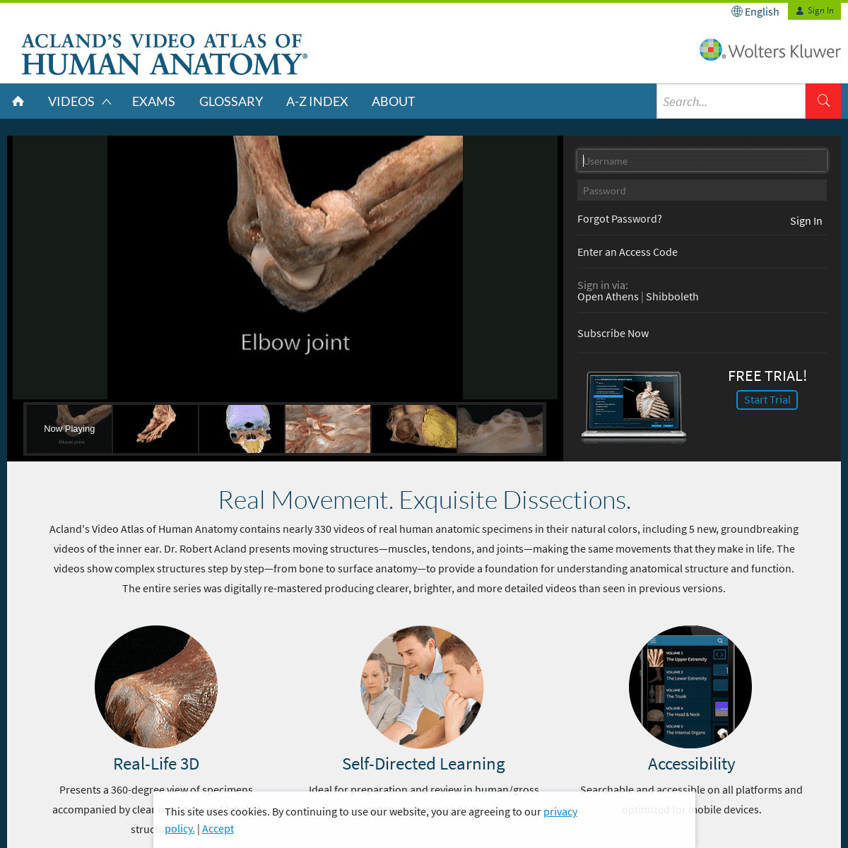
REAL MOVEMENT. EXQUISITE DISSECTIONS. Acland's Video Atlas of Human Anatomy contains nearly 330 videos of real human anatomic specimens in their natural colors, including 5 new, groundbreaking videos of the inner ear. Dr. Robert Acland presents moving structures—muscles, tendons, and joints—making the same movements that they make in life. The videos show complex structures step by step—from bone to surface anatomy—to provide a foundation for understanding anatomical structure and function. The entire series was digitally re-mastered producing clearer, brighter, and more detailed videos than seen in previous versions.REAL-LIFE 3D
Presents a 360-degree view of specimens accompanied by clear narration and labeled structures. SELF-DIRECTED LEARNING Ideal for preparation and review in human/gross anatomy courses andlabs.
ACCESSIBILITY
Searchable and accessible on all platforms and optimized for mobiledevices.
REVIEW AND EXAM PREP Interactive timed multiple-choice exams prepare students for practical exams, track their performance, and direct them back to correlatedvideos for review.
Start Your 48-Hour Free Trial WANT INFORMATION ABOUT INSTITUTIONAL PRICING? Contact us about group or site licensing for your school, hospital, orother institution.
Contact us
�2020 Wolters Kluwer Health, Inc. and/or its affiliates. All rightsreserved.
* HELP
* TERMS OF USE
* PRIVACY POLICY
* CONTACT US
* SUBSCRIPTIONS
* USER GUIDE
* Get Adobe Acrobat
� 2020 Wolters Kluwer Health, Inc. and/or its affiliates. All rightsreserved.
×
×
Enter an Access Code We are unable to redeem your access code. Please try again anothertime. Submit
Feedback Please take a moment to tell us about your experience withAclandAnatomy!
Please take a moment to tell us about your experience withAclandAnatomy!
(1000 characters left) Ease of use 1 = Not easy to use; 5 = Very easy to use1
2
3
4
5
Video navigation 1 = Not easy to navigate; 5 = Very easy to navigate1
2
3
4
5
Search results 1 = Not relevant; 5 = Very relevant1
2
3
4
5
Value to your understanding of the subject 1 = Not valuable; 5 = Veryvaluable
1
2
3
4
5
Do you currently use another format of the Acland product (DVDs, streaming/institutional version, etc.)?Yes
No
Tell us who you are.Student
Faculty
Professional
Other (Please specify) Other (Please specify) May we contact you about your feedback?Yes
No
Submit Feedback
Your feedback has been successfully submitted. We are unable to receive your feedback at this time. Please try againanother time.
Please sign in to submit feedback. Already a Subscriber? Sign InRequired Required
Forgot Password?
Enter an Access CodeSign in via:
Open Athens | Shibboleth×
This site uses cookies. By continuing to use our website, you are agreeing to our privacy policy.| Accept
Details
4